
Dostęp do tego artykułu jest płatny.
Zapraszamy do zakupu!
Po dokonaniu zakupu artykuł w postaci pliku PDF prześlemy bezpośrednio pod twój adres e-mail.
A Rare Case of Orthograde Retrieval of Extruded Instrument following Periapical Tissue Healing: Case Report
Abstract
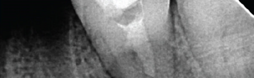
The ideal retrieval protocol of separated instruments reverts the case to the initial situation prior to the fracture incidence while preserving the tooth hard tissue and the integrity of the supporting structures. When a patient presented for emergency treatment of tooth #37 diagnosed with acute suppurative apical periodontitis, radiographic examination revealed a fractured instrument extruded into the periapex. The treatment options for retrieval were limited to replantation. The initial emergency treatment which consisted of orthograde pus drain, radicular disinfection, and intracanal calcium hydroxide dressing completely resolved patient’s symptoms. The follow-up radiographs revealed an interesting finding: gradual shift in the separated fragment positio into the radicular space allowing a successful nonsurgical removal of the broken instrument. In conclusion, the reaction of periodontal tissues to an extruded instrument fragment remaining in situ may be favourable; thus, a risk and benefit analysis approach is essential to fractured instrument retrieval.
References
- B. McGuigan, C. Louca, and H. F. Duncan, “Endodontic instrument fracture: causes and prevention,” British Dental Journal, vol. 214, no. 7, pp. 341–348, 2013.
- B. McGuigan, C. Louca, and H. F. Duncan, “Clinical decision-making after endodontic instrument fracture,” British Dental Journal, vol. 214, no. 8, pp. 395–400, 2013.
- Suter, A. Lussi, and P. Sequeira, “Probability of removing fractured instruments from root canals,” International Endodontic Journal, vol. 38, no. 2, pp. 112–123, 2005.
- Hülsmann and I. Schinkel, “Influence of several factors on the success or failure of removal of fractured instruments from the root canal,” Endodontics & Dental Traumatology, vol. 15, no. 6, pp. 252–258, 1999.
- B. McGuigan, C. Louca, and H. F. Duncan, “The impact of fractured endodontic instruments on treatment outcome,” British Dental Journal, vol. 214, no. 6, pp. 285–289, 2013.
- Panitvisai, P. Parunnit, C. Sathorn, and H. H. Messer, “Impact of a retained instrument on treatment outcome: a systematic review and meta-analysis,” Journal of Endodontia, vol. 36, no. 5, pp. 775–780, 2010.
- Spili, P. Parashos, and H. H. Messer, “The impact of instrument fracture on outcome of endodontic treatment,” Journal of Endodontia, vol. 31, no. 12, pp. 845–850, 2005.
- H. Kaddoura and A. A. Madarati, “Management of an overextruded fragment in a C-shaped root canal configuration: a case report and literature review,” Journal of Taibah University Medical Sciences, vol. 15, no. 5, pp. 431–436, 2020.
- Fujii, K. Morinaga, T. Asai et al., “Intentional replantation to treat apical periodontitis of maxillary first molar with foreign body located outside apical foramen using CBCT: a case report,” The Bulletin of Tokyo Dental College, vol. 61, no. 2, pp. 127–133, 2020.
- L. Satheesh, S. Jain, A. C. Bhuyan, and L. S. Devi, “Surgical management of a separated endodontic instrument using second generation platelet concentrate and hydroxyapatite,” Journal of Clinical and Diagnostic Research, vol. 11, no. 6, pp. ZD01–ZD03, 2017.
- Heydari, M. Rahmani, and M. Heydari, “Removal of a broken instrument from a tooth with apical periodontitis using a novel approach,” Iranian Endodontic Journal, vol. 11, no. 3, pp. 237–240, 2016.
- Plascencia, A. Cruz, R. Solís, M. Díaz, and J. Vázquez, “Iatrogenic displacement of a foreign body into the periapical tissues,” Case Reports in Dentistry, vol. 2014, Article ID 698538, 2014.
- Srivastava and N. Vineeta, “Foreign body in the periradicular area,” Journal of Endodontics, vol. 27, no. 9, pp. 593-594, 2001.
- Parashos and H. H. Messer, “Rotary NiTi instrument fracture and its consequences,” Journal of Endodontics, vol. 32, no. 11, pp. 1031–1043, 2006.
- Gao, Y. Shen, X. Zhou, and M. Haapasalo, “Remaining root dentin thickness in mesiobuccal canals of maxillary first molars after attempted removal of broken instrument fragments,” Australian Endodontic Journal, vol. 41, no. 3, pp. 122–127, 2015.
- A. Madarati, A. J. Qualtrough, and D. C. Watts, “Vertical fracture resistance of roots after ultrasonic removal of fractured instruments,” International Endodontic Journal, vol. 43, no. 5, pp. 424–429, 2010.
- D. Eleazer, “Lack of corrosion of stainless steel instruments in vivo by scanning electron microscope and microprobe analysis,” Journal of Endodontics, vol. 17, no. 7, pp. 346–349, 1991.
- Ryhänen, M. Kallioinen, J. Tuukkanen et al., “In vivo biocompatibility evaluation of nickel-titanium shape memory metal alloy: muscle and perineural tissue responses and encapsule membrane thickness,” Journal of Biomedical Materials Research, vol. 41, no. 3, pp. 481–488, 1998.
- Yusuf, “The significance of the presence of foreign material periapically as a cause of failure of root treatment,” Oral Surgery, Oral Medicine, and Oral Pathology, vol. 54, no. 5, pp. 566–574, 1982.
- N. Ramachandran Nair, “Non-microbial etiology: foreign body reaction maintaining post-treatment apical periodontitis,” Endodontic Topics, vol. 6, no. 1, pp 114–134, 2003.
- G. Khabbaz and P. D. Papadopoulos, “Deposition of calcified tissue around an overextended gutta-percha cone: case report,” International Endodontic Journal, vol. 32, no. 3, pp. 232–235, 1999.
- L. Smith, “The role of epithelium in the healing of experimental extraction wounds,” Journal of Dental Research, vol. 37, no. 2, pp. 187–194, 1958.