

Dostęp do tego artykułu jest płatny.
Zapraszamy do zakupu!
Po dokonaniu zakupu artykuł w postaci pliku PDF prześlemy bezpośrednio pod twój adres e-mail.
STUDIA PRZYPADKÓW
Torbiel kanału przysiecznego – diagnostyka i leczenie
Incisive canal cyst – diagnosis and treatment
Paulina Adamska, Kosma Mazur, Dorota Pylińska-Dąbrowska, Natalia Kobusińska, Adam Zedler
Streszczenie
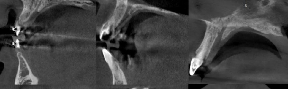
Torbiel kanału przysiecznego to jedna z najczęstszych torbieli w obrębie kości szczęk. Zmiana patologiczna jest wykrywana przypadkowo, podczas rutynowych badań radiologicznych lub w związku z objawami, takimi jak rozdęcie wyrostka czy dolegliwości bólowe, lub w przypadku zakażenia i zropienia. W niniejszej pracy przedstawiono metody diagnostyki i leczenia pacjentów z torbielą kanału przysiecznego.
Abstract
The incisive canal cyst is one of the most common cysts in the jaw bones. The lesion is found incidentally during routine radiological examinations or in association with symptoms such as distension of the alveolar process, pain or in case of infection and purulence. This study presents methods of diagnosis and treatment of patients with incisive canal cysts.
Hasła indeksowe: torbiel kanału przysiecznego, wyłyżeczkowanie torbieli, CBCT, tomografia komputerowa wiązki stożkowej
Key words: incisive canal cyst, cyst curettage, CBCT, cone beam computed tomography
Piśmiennictwo
1. Lake S, Iwanaga J, Kikuta S i wsp. The incisive canal. A comprehensive review. Cureus. 2018; 10(7): e3069.
2. Jacob S, Zelano B, Gungor A i wsp. Location and gross morphology of the nasopalatine duct in human adults. Arch Otolaryngol Head Neck Surg. 2000; 126(6): 741-748.
3. Miwa Y, Asaumi R, Kawai T i wsp. Morphological observation and CBCT of the bony canal structure of the groove and the location of blood vessels and nerves in the palatine of elderly human cadavers. Surg Radiol Anat. 2018; 40(2): 199-206.
4. Salemi F, Moghadam FA, Shakibai Z i wsp. Three-dimensional assessment of the nasopalatine canal and the surrounding bone using cone-beam computed tomography. J Periodontol Implant Dent. 2016; 8(1): 1-7.
5. Khojastepour L, Haghnegahdar A, Keshktar M. Morphology and dimensions of nasopalatine canal. A radiographic analysis using cone beam computed tomography. J Dent (Shiraz). 2017; 18(4): 244-250.
6. Güncü GN, Yıldırım YD, Yılmaz HG i wsp. Is there a gender difference in anatomic features of incisive canal and maxillary environmental bone? Clin Oral Implants Res. 2013; 24(9): 1023-1026.
7. Al-Amery SM, Nambiar P, Jamaludin M i wsp. Cone beam computed tomography assessment of the maxillary incisive canal and foramen. Considerations of anatomical variations when placing immediate implants. PLoS One. 2015; 10(2): e0117251.
8. Bahşi I, Orhan M, Kervancıoğlu P i wsp. Anatomical evaluation of nasopalatine canal on cone beam computed tomography images. Folia Morphol (Warsz). 2019; 78(1): 153-162.
9. McCrea SJ. Nasopalatine duct cyst, a delayed complication to successful dental implant placement. Diagnosis and surgical management. J Oral Implantol. 2014; 40(2): 189-195.
10. Cecchetti F, Ottria L, Bartuli F i wsp. Prevalence, distribution, and differential diagnosis of nasopalatine duct cysts. Oral Implantol (Rome). 2012; 5(2-3): 47-53.
11. Dedhia P, Dedhia S, Dhokar A i wsp. Nasopalatine duct cyst. Case Rep Dent. 2013; 2013: 869516.
12. Ueda N, Tanaka T, Oda M i wsp. Advocacy of diagnostic criteria for maxillary incisive canal cysts based on alteration of normal maxillary incisive canals according to aging in Japanese populations. Head Face Med. 2019; 15(1): 25.
13. Suter VG, Jacobs R, Brücker MR i wsp. Evaluation of a possible association between a history of dentoalveolar injury and the shape and size of the nasopalatine canal. Clin Oral Investig. 2016; 20(3): 553-561.
14. Kaczmarzyk T. Torbiele obszaru szczękowo-twarzowego. Warszawa: Kwintesencja; 2015: 74.
15. Yeom HG, Kang JH, Yun SU i wsp. Nasopalatine duct cyst with sebaceous differentiation. A rare case report with literature review. BMC Oral Health. 2021; 21(1): 419.
16. Lang MJ, Lee YP, Hwang MJ i wsp. Nasopalatine duct cyst – case report. J Dent Sci. 2021; 16(3): 1047-1049.


