

Dostęp do tego artykułu jest płatny.
Zapraszamy do zakupu!
Cena: 24.00 PLN (z VAT)
Kup artykuł
Po dokonaniu zakupu artykuł w postaci pliku PDF prześlemy bezpośrednio pod twój adres e-mail.
MS 2022; 5: 16-24.
Zastosowanie wielofalowego lasera diodowego do usunięcia tłuszczaka techniką zachowania błony śluzowej
The use of multiwave diode laser for the removal of lipoma by using mucosal preservation technique
Marcin Tkaczyk, Rafał Wiench, Natalia Stefanik, Katarzyna Latusek, Aleksandra Warakomska, Dariusz Skaba
Streszczenie
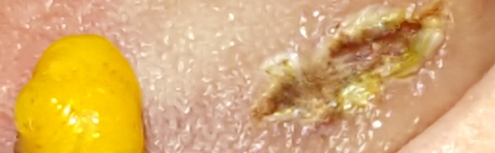
Tłuszczak jest częstym wolno rosnącym, łagodnym nowotworem, wywodzącym się z tkanki łącznej, zbudowanym z adipocytów. Dokładna przyczyna powstawania nie jest znana. W etiologii rozważa się: uraz, czynnik genetyczny oraz choroby współistniejące. Do metod leczenia należą: ostrzykiwanie preparatami kortykosteroidowymi, liposukcja oraz usunięcie chirurgiczne, które jest złotym standardem. Warta rozważenia alternatywa w stosunku do tradycyjnego chirurgicznego usunięcia tłuszczaka to zastosowanie laseroterapii. Celem artykułu jest prezentacja techniki zachowania błony śluzowej (MPT) jako małoinwazyjnego zabiegu chirurgicznego, zastosowana do usunięcia tłuszczaka zlokalizowanego na języku u 57-letniej, ogólnie zdrowej pacjentki. Zabieg przeprowadzono laserem diodowym 980 nm (moc średnia: 4W, tryb modulowany pracy, czas impulsu: 100 μs, czas przerwy: 100 μs, wypełnienie 50% światłowodem 320 µm metodą kontaktową). Po zabiegu wycięcia guza, aby przyspieszyć gojenie rany, zastosowano cykl fotobiomodulacji laserem 635 nm (moc wyjściowa: 100 mW, fluencja: 3 J/cm2, tryb pracy: CW, czas pojedynczej irradiacji: 30 s) z użyciem aplikatora szklanego 8 mm metodą kontaktową. Prezentowana technika MPT wykonywana laserem jest szybką i ergonomiczną alternatywą dla tradycyjnego chirurgicznego usunięcia skalpelem. Zapewnia warstwowe cięcie tkanek miękkich z jednoczasową hemostazą, dobry (niezaburzony krwawieniem) wgląd w pole zabiegowe, możliwość zachowania dużej ilości tkanek sklepienia, dzięki czemu nie ma konieczności zszywania rany.
Abstract
A lipoma is a common, slow-growing, benign tumor that originates in connective tissue and is made up of fat cells (adipocytes). Most often it is located on the trunk and is painless. The exact cause of formation is unknown. The etiology takes into account the trauma, genetic factors and coexisting diseases. In order to diagnose lipoma lesions, we use X-ray, MRI, USG and PET. Treatment methods include corticosteroid injection, liposuction and surgical removal, which is the gold standard. An alternative to traditional surgical removal of a lipoma that is worth considering is the use of laser therapy. The aim of this article is to present the mucosal preservation technique (MPT) as a minimally invasive surgical procedure to remove the lipoma located on the tongue in a 57-year-old, generally healthy patient. The treatment was performed with a 980 nm diode laser (average power: 4W, modulated mode, pulse time: 100 μs, pause time: 100 μs, 50% duty cycle, 320 µm optical fiber, contact method). After the tumor excision, in order to accelerate the wound healing, a photobiomodulation cycle was used with a 635 nm laser (output power: 100 mW, fluence: 3J/cm2, operating mode: CW, time of single irradiation: 30 s) with the use of an 8 mm glass applicator with the contact method. The presented MPT technique, performed with a laser, is a quick and ergonomic alternative to traditional surgical removal with a scalpel. Provides layered incision of soft tissues with simultaneous hemostasis, good (undisturbed bleeding) view of the treatment area, the possibility of preserving a large amount of surrounding tissues, thanks to which there is no necessary to suture the wound.
Hasła indeksowe: guzy łagodne, tłuszczak, laser diodowy, technika zachowania błony śluzowej, fotobiomodulacja
Key words: benign tumors, lipoma, diode laser, mucosal preservation technique, photobiomodulation
The use of multiwave diode laser for the removal of lipoma by using mucosal preservation technique
Marcin Tkaczyk, Rafał Wiench, Natalia Stefanik, Katarzyna Latusek, Aleksandra Warakomska, Dariusz Skaba
Streszczenie
Tłuszczak jest częstym wolno rosnącym, łagodnym nowotworem, wywodzącym się z tkanki łącznej, zbudowanym z adipocytów. Dokładna przyczyna powstawania nie jest znana. W etiologii rozważa się: uraz, czynnik genetyczny oraz choroby współistniejące. Do metod leczenia należą: ostrzykiwanie preparatami kortykosteroidowymi, liposukcja oraz usunięcie chirurgiczne, które jest złotym standardem. Warta rozważenia alternatywa w stosunku do tradycyjnego chirurgicznego usunięcia tłuszczaka to zastosowanie laseroterapii. Celem artykułu jest prezentacja techniki zachowania błony śluzowej (MPT) jako małoinwazyjnego zabiegu chirurgicznego, zastosowana do usunięcia tłuszczaka zlokalizowanego na języku u 57-letniej, ogólnie zdrowej pacjentki. Zabieg przeprowadzono laserem diodowym 980 nm (moc średnia: 4W, tryb modulowany pracy, czas impulsu: 100 μs, czas przerwy: 100 μs, wypełnienie 50% światłowodem 320 µm metodą kontaktową). Po zabiegu wycięcia guza, aby przyspieszyć gojenie rany, zastosowano cykl fotobiomodulacji laserem 635 nm (moc wyjściowa: 100 mW, fluencja: 3 J/cm2, tryb pracy: CW, czas pojedynczej irradiacji: 30 s) z użyciem aplikatora szklanego 8 mm metodą kontaktową. Prezentowana technika MPT wykonywana laserem jest szybką i ergonomiczną alternatywą dla tradycyjnego chirurgicznego usunięcia skalpelem. Zapewnia warstwowe cięcie tkanek miękkich z jednoczasową hemostazą, dobry (niezaburzony krwawieniem) wgląd w pole zabiegowe, możliwość zachowania dużej ilości tkanek sklepienia, dzięki czemu nie ma konieczności zszywania rany.
Abstract
A lipoma is a common, slow-growing, benign tumor that originates in connective tissue and is made up of fat cells (adipocytes). Most often it is located on the trunk and is painless. The exact cause of formation is unknown. The etiology takes into account the trauma, genetic factors and coexisting diseases. In order to diagnose lipoma lesions, we use X-ray, MRI, USG and PET. Treatment methods include corticosteroid injection, liposuction and surgical removal, which is the gold standard. An alternative to traditional surgical removal of a lipoma that is worth considering is the use of laser therapy. The aim of this article is to present the mucosal preservation technique (MPT) as a minimally invasive surgical procedure to remove the lipoma located on the tongue in a 57-year-old, generally healthy patient. The treatment was performed with a 980 nm diode laser (average power: 4W, modulated mode, pulse time: 100 μs, pause time: 100 μs, 50% duty cycle, 320 µm optical fiber, contact method). After the tumor excision, in order to accelerate the wound healing, a photobiomodulation cycle was used with a 635 nm laser (output power: 100 mW, fluence: 3J/cm2, operating mode: CW, time of single irradiation: 30 s) with the use of an 8 mm glass applicator with the contact method. The presented MPT technique, performed with a laser, is a quick and ergonomic alternative to traditional surgical removal with a scalpel. Provides layered incision of soft tissues with simultaneous hemostasis, good (undisturbed bleeding) view of the treatment area, the possibility of preserving a large amount of surrounding tissues, thanks to which there is no necessary to suture the wound.
Hasła indeksowe: guzy łagodne, tłuszczak, laser diodowy, technika zachowania błony śluzowej, fotobiomodulacja
Key words: benign tumors, lipoma, diode laser, mucosal preservation technique, photobiomodulation
PIŚMIENNICTWO
- Weiss SW. Lipomatous tumors. Monogr Pathol. 1996; 38: 207-239.
- Neville BW, Damm DD, Allen CM i wsp. Color atlas of oral and maxillofacial diseases [e-book]. Elsevier Health Sciences. 2018; 12: 318.
- Vásquez Elera L, Guzman Rojas P, Sánchez Herrera M i wsp. [Familiar adenomatous polyposis: report of 2 cases]. Rev Gastroenterol Peru. 2018; 38(1): 78-81.
- Aust MC, Spies M, Kall S i wsp. Posttraumatic lipoma: fact or fiction? Skinmed. 2007; 6(6): 266-270.
- Barisella M, Giannini L, Piazza C. From head and neck lipoma to liposarcoma. A wide spectrum of differential diagnoses and their therapeutic implications. Curr Opin Otolaryngol Head Neck Surg. 2020; 28(2): 136-143.
- Panagopoulos I, Gorunova L, Agostini A i wsp. Fusion of the HMGA2 and C9orf92 genes in myolipoma with t(9;12)(p22;q14). Diagn Pathol. 2016; 11: 22.
- Demicco EG. Molecular updates in adipocytic neoplasms✰. Semin Diagn Pathol. 2019; 36(2): 85-94.
- Forcucci JA, Sugianto JZ, Wolff DJ i wsp. „Low-fat” pseudoangiomatous spindle cell lipoma. A rare variant with loss of 13q14 region. Am J Dermatopathol. 2015; 37(12): 920-923.
- Brasfield RD, Das Gupta TK. Liposarcoma. CA Cancer J Clin. 1970; 20(1): 3-8.
- Katsuyama Y, Shirai T, Terauchi R i wsp. Chondroid lipoma of the neck. A case report. BMC Res Notes. 2018; 11(1): 415.
- Johnson CN, Ha AS, Chen E i wsp. Lipomatous soft-tissue tumors. J Am Acad Orthop Surg. 2018; 26(22): 779-788.
- Salam GA. Lipoma excision. Am Fam Physician. 2002; 65(5): 901-904.
- Silistreli OK, Durmuş EU, Ulusal BG i wsp. What should be the treatment modality in giant cutaneous lipomas? Review of the literature and report of 4 cases. Br J Plast Surg. 2005; 58(3): 394-398.
- Romeo U, Palaia G, Tenore G i wsp. Excision of oral mucocele by different wavelength lasers. Indian J Dent Res. 2013; 24(2): 211-215.
- Kopp WK, St-Hilaire H. Mucosal preservation in the treatment of mucocele with CO2 laser. J Oral Maxillofac Surg. 2004; 62(12): 1559-1561.
- Huang IY, Chen CM, Kao YH i wsp. Treatment of mucocele of the lower lip with carbon dioxide laser. J Oral Maxillofac Surg. 2007; 65(5): 855-858.
- Sidorowicz K, Błochowiak K, Sokalski J. Zastosowanie lasera Er:YAG w zabiegach chirurgii stomatologicznej. Dental Forum. 2014; XLII(1): 87-92.


