

Dostęp do tego artykułu jest płatny.
Zapraszamy do zakupu!
Cena: 12.50 PLN (z VAT)
Kup artykuł
Po dokonaniu zakupu artykuł w postaci pliku PDF prześlemy bezpośrednio pod twój adres e-mail.
MS 2021; 6: 60-68.
Zastosowanie Cariogramu w ocenie ryzyka próchnicy u pacjentek z cukrzycą
Joanna Kunert, Monika Colonna‑Walewska, Elżbieta Bołtacz‑Rzepkowska
Streszczenie
Wprowadzenie. Określenie indywidualnego profilu ryzyka próchnicy to kluczowy element przy planowaniu postępowania profilaktyczno‑leczniczego. Istotą sukcesu w kwalifikacji pacjenta do grupy ryzyka jest identyfikacja czynników mających wpływ na progresję próchnicy.
Cel pracy. Ocena ryzyka próchnicy u pacjentek chorujących na cukrzycę, z wykorzystaniem programu Cariogram.
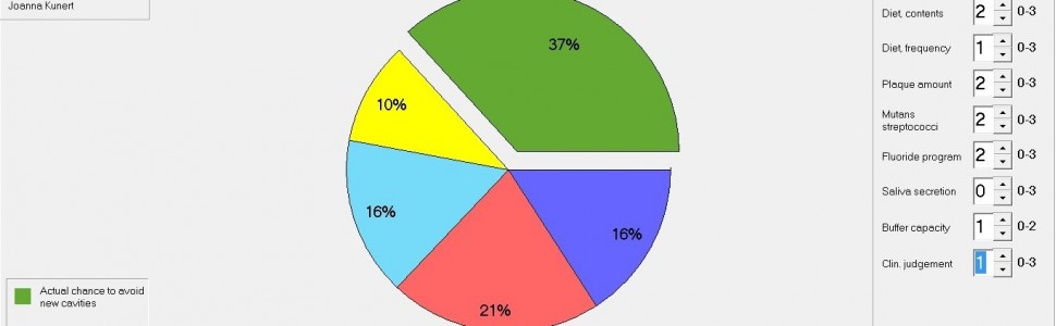
Materiał i metody. Badaniem objęto 52 pacjentki Poradni Diabetologicznej w Łodzi, chore na cukrzycę insulinozależną – typu 1 (40 z cukrzycą niewyrównaną i 12 z wyrównaną). Grupę porównawczą stanowiły 52 pacjentki bez cukrzycy. Oceny ryzyka próchnicy dokonano na podstawie programu Cariogram, w którym każdemu z czynników ryzyka próchnicy przypisuje się wartości liczbowe w zależności od nasilenia cechy. Program po przetworzeniu danych przedstawia diagram kołowy, obrazujący procentowy udział poszczególnych czynników w progresji próchnicy. Wraz z obniżaniem się wartości wskaźnika wzrasta ryzyko wystąpienia próchnicy.
Wyniki. Wartość Cariogramu wśród pacjentek zdrowych wyniosła 72%, a u pacjentek z cukrzycą – 37%. Wnioski. Pacjentki chore na cukrzycę stanowią grupę zwiększonego ryzyka występowania próchnicy zębów i dlatego wymagają szczególnej opieki stomatologicznej. Cariogram jest skutecznym narzędziem nie tylko do oceny ryzyka próchnicy, lecz także do planowania działań profilaktyczno‑leczniczych.
Abstract
Introduction. Determining the individual caries risk profile is a key element in planning preventive and therapeutic procedure. The essence of success in qualifying a patient to a risk group is the identification of factors influencing the progression of caries.
Aim of the study. Caries risk assessment in diabetic patients using the Cariogram program.
Material and method. The study included 52 patients of the Diabetes Clinic in Łódź, with insulin‑dependent diabetes mellitus‑type 1 (40 with uncontrolled diabetes and 12 with compensated diabetes). The comparative group consisted of 52 nondiabetic patients. Caries risk assessment was based on Cariogram program, in which each of the caries risk factors are assigned numerical values depending on the severity of the features. After processing the data, the program presents a pie diagram showing the percentage share of individual factors in the caries progression. As the index value decreases, the risk of caries increases.
Results. The value of the Cariogram among healthy patients was 72%, and in patients with diabetes‑37%.
Conclusions. Diabetic patients constitute a group of increased risk of tooth decay and therefore require special dental care. The cariogram is an effective tool not only for caries risk assessment, but also for planning preventive and therapeutic measures.
Hasła indeksowe: ryzyko próchnicy, cukrzyca, Cariogram
Key words: caries risk, diabetes, Cariogram
Piśmiennictwo
- World Health Organization. Classification of diabetes mellitus. Geneva; 2019.
- Bąk E, Nowak‑Kapusta Z, Dobrzyn‑Matusiak D i wsp. An assessment of diabetes‑dependent quality of life (ADDQoL) in women and men in Poland with type 1 and type 2 diabetes. Ann Agric Environ Med. 2019; 26(3): 429‑438.
- Mauri‑Obradors E, Estrugo‑Devesa A, Jané‑Salas E i wsp. Oral manifestations of diabetes mellitus. A systematic review. Med Oral Patol Oral Cir Bucal. 2017; 22(5): e586‑e594.
- Leite RS, Marlow NM, Fernandes JK i wsp. Oral health and type 2 diabetes. Am J Med Sci. 2013; 345(4): 271‑273.
- Lalla E, Papapanou PN. Diabetes mellitus and periodontitis: a tale of two common interrelated diseases. Nat Rev Endocrinol. 2011; 7(12): 738‑748.
- López R, Smith PC, Göstemeyer G i wsp. Ageing, dental caries and periodontal diseases. J Clin Periodontol. 2017; 44(Suppl 18): S145‑S152.
- Majbauddin A, Tanimura C, Aoto H i wsp. Association between dental caries indicators and serum glycated hemoglobin‑levels among patients with type 2 diabetes mellitus. J Oral Sci. 2019; 61(2): 335‑342.
- Almusawi MA, Gosadi I, Abidia R i wsp. Potential risk factors for dental caries in type 2 diabetic patients. Int J Dent Hyg. 2018; 16(4): 467‑475.
- Machiulskiene V, Campus G, Carvalho JC i wsp. Terminology of Dental Caries and Dental Caries Management. Consensus Report of a Workshop Organized by ORCA and Cariology Research Group of IADR. Caries Res. 2020; 54(1): 7‑14.
- Rafatjou R, Razavi Z, Tayebi S i wsp. Dental health status and hygiene in children and adolescents with type 1 diabetes mellitus. J Res Health Sci. 2016; 16(3): 122‑126.
- Bali C, Gurdet C, Hauer G i wsp. Evaluation of the effect of professional dental cleaning and education in dental hygiene in type 1 diabetic patients. Acta Med Austriaca. 1999; 26(5): 159‑162.
- Pérez‑Losada FL, Jané‑Salas E, Sabater‑Recolons MM i wsp. Correlation between periodontal disease management and metabolic control of type 2 diabetes mellitus. A systematic literature review. Med Oral Patol Oral Cir Bucal. 2016; 21(4): e440‑e446.
- Oberti L, Gabrione F, Nardone M i wsp. Two‑way relationship between diabetes and periodontal disease. A reality or a paradigm? J Biol Regul Homeost Agents. 2019; 33(3 Suppl 1): 153‑159.
- Jaedicke KM, Bissett SM, Finch T i wsp. Exploring changes in oral hygiene behaviour in patients with diabetes and periodontal disease. A feasibility study. Int J Dent Hyg. 2019; 17(1): 55‑63.
- Hänsel Petersson G. Assessing caries risk – using the Cariogram model. Swed Dent J Suppl. 2003; 158: 1‑65.
- Bratthall D, Hänsel Petersson G. Cariogram – a multifactorial risk assessment model for a multifactorial disease. Community Dent Oral Epidemiol. 2005; 33(4): 256‑264.


