MS 2021; 5: 26-31.
Leczenie postaci zanikowo-nadżerkowej liszaja płaskiego jamy ustnej z użyciem lasera. Opis przypadku
Kamila Kozak-Jastrzębska, Elżbieta Dembowska, Renata Majka, Urszula Kiczka, Szymon Gacek
Streszczenie
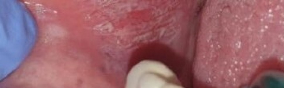
Liszaj płaski jamy ustnej (oral lichen planus – OLP) jest przewlekłą zapalną chorobą błony śluzowej związaną z dysfunkcją komórek immunokompetentnych. Jego manifestacją kliniczną mogą być białe zmiany (w postaciach siateczkowej i płytkowej OLP) oraz czerwone zmiany(w postaciach: zanikowej, pęcherzowej oraz nadżerkowej). Zmiany erozyjne, atroficzne i wrzodziejące wymagają długotrwałego leczenia ogólnego w związku z towarzyszącym im zapaleniem i silnym bólem. Etiologia liszaja płaskiego nie została dokładnie poznana, a skuteczne leczenie jest bardzo trudne. Na podstawie aktualnych doniesień z piśmiennictwa przedstawiono możliwości leczenia laserowego laserem diodowym 980 nm jednej z częściej spotykanych postaci OLP –zanikowo-nadżerkowej. Leczenie to jest w większości przypadków objawowe, ze względu na nawrotowy charakter zmian. W pracy przedstawiono dokładny protokół leczenia i jego efekty kliniczne. Laser diodowy 980 nm ułatwia gojenie się zmian, zmniejsza objawy zapalenia i objawy bólowe, co wpływa na poprawę jakości życia pacjentów. Polecane jest leczenie miejscowe wykwitów liszaja płaskiego przed wprowadzeniem leczenia ogólnoustrojowego z zastosowaniem leków glikokortykosteroidowych.
Abstract
Oral lichen planus (OLP) is a chronic mucosal disease associated with immunocompetent cell dysfunction. Clinically, it can occur in the form of white lesions: in reticular and lamellar types, and in the form of red lesions, present in atrophic, blistering and erosional types. Erosive, atrophic and ulcerative lesions require long-term general treatment for inflammation and the accompanying severe pain. The aetiology of lichen planus is not exactly known and effective treatment is very difficult. On the basis of current literature reports, the possibilities of 980 nm diode laser treatment with one of the most common forms of OLP – atrophic-erosional form – were presented. This treatment in most cases is symptomatic, caused by the recurring nature of the changes. The paper presents a detailed treatment protocol and its effects. The 980 nm diode laser facilitates the healing of lesions, reduces the symptoms of inflammation and clinical symptoms, which improves patients’ quality of life. Topical treatment of lichen planus lesions is recommended due to the occurrence of minor side effects of this therapy, before we decided to use systemic steroids treatment.
Hasła indeksowe: liszaj płaski jamy ustnej, laser diodowy 980 nm
Key words: oral lichen planus, diode laser 980 nm
PIŚMIENNICTWO
1. Bigby M. The relationship between lichen planus and hepatitis C clarified. Arch Dermatol. 2009; 145: 1048-50.
2. Monteiro BV, Pereira Jdos S, Nonaka CF. Immunoexpression of Th17-related cytokines in oral lichen planus. Appl Immunohistochem Mol Morphol. 2015; 23: 409–415.
3. Bermejo-Fenoll A, Sánchez-Siles M, López-Jornet P i wsp. A retrospective clinicopathological study of 550 patients with oral lichen planus in south-eastern Spain. J oral Pathol Med. 2010; 39: 491-6.
4. He Y, Gong D, Shi C i wsp. Dysbiosis of oral buccal mucosa microbiota in patients with oral lichen planus. [J] Oral Dis. 2017; 23(5): 674–682.
5. Azma E, Safavi N. Diode laser application in softtissueoralsurgery. J Lasers Med Sci. 2013; 4(4).
6. Carbone M, Arduinop G, Carrozzo M et al. Course of oral lichen planus: a retrospective study of 808 northern italian patients. Oral Dis. 2009; 15: 235-43.
7. Gorouhi F, Davari PN. Cutaneous and Mucosal Lichen Planus: A Comprehensive Review of Clinical Subtypes, Risk Factors, Diagnosis, and Prognosis. Scientific World Journal. 2014, 1-22.
8. Thongprasom K, Carrozzo M, Furness S, Lodi G. Interventions for treating oral lichen planus. Cochrane Database Syst Rev. 2011; 7:CD001168.
9. Kamath VV, Setlur K, Yerlagudda K. Oral lichenoid lesions – a review and update. Indian J Dermatol. 2015; 60: 102.
10. Lomke MA. Clinical applications of dental lasers. Gen Dent. 2009; 57(1).