

Dostęp do tego artykułu jest płatny.
Zapraszamy do zakupu!
Cena: 12.50 PLN (z VAT)
Kup artykuł
Po dokonaniu zakupu artykuł w postaci pliku PDF prześlemy bezpośrednio pod twój adres e-mail.
Multiple peripheral mandible osteomas – a case report
Tomasz Barłóg
Streszczenie
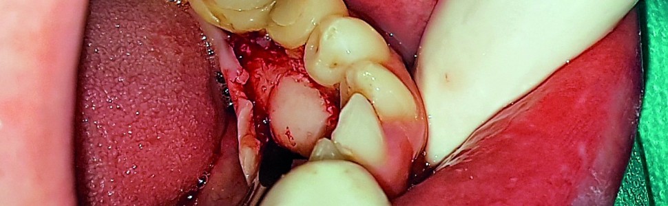
Kostniaki są łagodnymi guzami o niskiej dynamice wzrostu, zbudowanymi z dojrzałej tkanki kostnej zbitej lub gąbczastej. W postaci mnogiej mogą stanowić objaw zespołu Gardnera. W zależności od lokalizacji dzieli się je na centralne, obwodowe i pozaszkieletowe. Kostniaki mogą lokalizować się w różnych miejscach w obrębie głowy. Ich przebieg kliniczny często jest asymptomatyczny. W niektórych przypadkach powodują defekty estetyczne i różnorodne dysfunkcje i z tego powodu wymagają interwencji chirurgicznej. Celem pracy było przedstawienie przypadku pacjenta z mnogimi kostniakami obwodowymi żuchwy. Zmiany radiologicznie dawały obraz jednolicie silnie cieniujących mas o wyraźnie zaznaczonych brzegach. Histologicznie przypominały dojrzałą kość kortykalną.
Abstract
Osteomas are benign lesions with low dynamics of growth, composed of mature compact or spongy bone. In case of multifocal form they may be a symptom of Gardner’s syndrome. Depends on location they are classified as central, peripheral or extraskeletal. They may be situated at different parts of a head. Clinical course is often asymptomatic. In some instances they cause esthetic defects and functional disorders, thus require surgical intervention. The purpose of this article was to present a case of multiple peripheral mandible osteomas. The lesions displayed an image of homogenous, radiopaque, well-defined mass. Histologically they resembled mature cortical bone.
Hasła indeksowe: kostniak, egzostoza, zespół Gardnera, guzy żuchwy
Key words: osteoma, exostosis, Gardner’s syndrome, mandible tumors
PIŚMIENNICTWO
1. Borghesi A. i wsp.: Peripheral osteoma, compound odontoma, focal cemento-osseous dysplasia, and cemento-ossifying fibroma in the same hemimandible: CBCT findings of an unusual case. Radiol. Case Rep., 2017, 12, 4, 756-759.
2. Kim S.H. i wsp.: Post-traumatic peripheral giant osteoma in the frontal bone. Arch. Craniofac. Surg., 2017, 18, 4, 273-276.
3. Savas S.A. i wsp.: Outer side of the nasal bone osteoma. J. Craniofac. Surg., 2017, 28, 4, e399-e400.
4. Karataş A. i wsp.: Osteoma originating from mastoid cortex. Turk. Arch. Otorhinolaryngol., 2017, 55, 1, 48-50.
5. Ishii T. i wsp.: A giant osteoma of the ethmoid sinus. J. Craniofac. Surg., 2018, 29, 3, 661-662.
6. Viswanatha B.: Maxillary sinus osteoma: two cases and review of the literature. Acta Otorhinolaryngol. Ital., 2012, 32, 3, 202-205.
7. Tempaku A.: Multiple skull osteomas in a 24‐year‐old woman. J. Gen. Fam. Med., 2017, 18, 6, 468-469.
8. Bai W.L. i wsp.: Removal of a giant ethmoidal sinus osteoma with orbital extension. J. Clin. Otorhinolaryngol. Head Neck Surg., 2017, 31, 24, 1932-1934.
9. Yang Y., Wang L., Wu Z.: Frontal sinus osteoma accompanied by intracranial mucocele and local hyperostosis frontalis interna. World Neurosurg., 2018, 113, 94-95.
10. de Souza N.T. i wsp.: An unusual osteoma in the mandibular condyle and the successful replacement of the temporomandibular joint with a custom-made prosthesis: a case report. BMC Res. Notes, 2017, 10, 1, 727.
11. Iwai T. i wsp.: Peripheral osteoma of the mandibular notch: report of a case. Iran. J. Radiol., 2013, 10, 2, 74-76.
12. Vashishth S. i wsp.: An unusual cause for trismus caused by mandibular coronoidosteoma: a case report. Imaging Sci. Dent., 2013, 43, 1, 45-48.
13. Soni S. i wsp.: Revisiting peripheral osteoma of the mandible with case series and review of literature. Indian J. Otolaryngol. Head Neck Surg., 2014, 66, 2, 212-218.
14. Ragupathy K. i wsp.: Peripheral osteoma of the body of mandible: a case report. J. Maxillofac. Oral Surg., 2015, 14, 4, 1004-1008.
15. Nilesh K. i wsp.: Solitary peripheral ivory osteoma of the mandible presenting with difficulty in deglutition: a case report. J. Dent. Res. Dent. Clin. Dent. Prospects, 2017, 11, 1, 56-60.
16. Tarsitano A. i wsp.: Unusual presentation of obstructive sleep apnoea syndrome due to a giant mandible osteoma: case report and literature review. Acta Otorhinolaryngol. Ital., 2013, 33, 1, 63-66.
17. Kaczmarzyk T. i wsp.: Nowotwory zębopochodne i guzy nowotworopodobne kości szczękowych. Wydawnictwo Kwintesencja, Warszawa 2009, 35, 83, 235.
Tomasz Barłóg
Streszczenie
Kostniaki są łagodnymi guzami o niskiej dynamice wzrostu, zbudowanymi z dojrzałej tkanki kostnej zbitej lub gąbczastej. W postaci mnogiej mogą stanowić objaw zespołu Gardnera. W zależności od lokalizacji dzieli się je na centralne, obwodowe i pozaszkieletowe. Kostniaki mogą lokalizować się w różnych miejscach w obrębie głowy. Ich przebieg kliniczny często jest asymptomatyczny. W niektórych przypadkach powodują defekty estetyczne i różnorodne dysfunkcje i z tego powodu wymagają interwencji chirurgicznej. Celem pracy było przedstawienie przypadku pacjenta z mnogimi kostniakami obwodowymi żuchwy. Zmiany radiologicznie dawały obraz jednolicie silnie cieniujących mas o wyraźnie zaznaczonych brzegach. Histologicznie przypominały dojrzałą kość kortykalną.
Abstract
Osteomas are benign lesions with low dynamics of growth, composed of mature compact or spongy bone. In case of multifocal form they may be a symptom of Gardner’s syndrome. Depends on location they are classified as central, peripheral or extraskeletal. They may be situated at different parts of a head. Clinical course is often asymptomatic. In some instances they cause esthetic defects and functional disorders, thus require surgical intervention. The purpose of this article was to present a case of multiple peripheral mandible osteomas. The lesions displayed an image of homogenous, radiopaque, well-defined mass. Histologically they resembled mature cortical bone.
Hasła indeksowe: kostniak, egzostoza, zespół Gardnera, guzy żuchwy
Key words: osteoma, exostosis, Gardner’s syndrome, mandible tumors
PIŚMIENNICTWO
1. Borghesi A. i wsp.: Peripheral osteoma, compound odontoma, focal cemento-osseous dysplasia, and cemento-ossifying fibroma in the same hemimandible: CBCT findings of an unusual case. Radiol. Case Rep., 2017, 12, 4, 756-759.
2. Kim S.H. i wsp.: Post-traumatic peripheral giant osteoma in the frontal bone. Arch. Craniofac. Surg., 2017, 18, 4, 273-276.
3. Savas S.A. i wsp.: Outer side of the nasal bone osteoma. J. Craniofac. Surg., 2017, 28, 4, e399-e400.
4. Karataş A. i wsp.: Osteoma originating from mastoid cortex. Turk. Arch. Otorhinolaryngol., 2017, 55, 1, 48-50.
5. Ishii T. i wsp.: A giant osteoma of the ethmoid sinus. J. Craniofac. Surg., 2018, 29, 3, 661-662.
6. Viswanatha B.: Maxillary sinus osteoma: two cases and review of the literature. Acta Otorhinolaryngol. Ital., 2012, 32, 3, 202-205.
7. Tempaku A.: Multiple skull osteomas in a 24‐year‐old woman. J. Gen. Fam. Med., 2017, 18, 6, 468-469.
8. Bai W.L. i wsp.: Removal of a giant ethmoidal sinus osteoma with orbital extension. J. Clin. Otorhinolaryngol. Head Neck Surg., 2017, 31, 24, 1932-1934.
9. Yang Y., Wang L., Wu Z.: Frontal sinus osteoma accompanied by intracranial mucocele and local hyperostosis frontalis interna. World Neurosurg., 2018, 113, 94-95.
10. de Souza N.T. i wsp.: An unusual osteoma in the mandibular condyle and the successful replacement of the temporomandibular joint with a custom-made prosthesis: a case report. BMC Res. Notes, 2017, 10, 1, 727.
11. Iwai T. i wsp.: Peripheral osteoma of the mandibular notch: report of a case. Iran. J. Radiol., 2013, 10, 2, 74-76.
12. Vashishth S. i wsp.: An unusual cause for trismus caused by mandibular coronoidosteoma: a case report. Imaging Sci. Dent., 2013, 43, 1, 45-48.
13. Soni S. i wsp.: Revisiting peripheral osteoma of the mandible with case series and review of literature. Indian J. Otolaryngol. Head Neck Surg., 2014, 66, 2, 212-218.
14. Ragupathy K. i wsp.: Peripheral osteoma of the body of mandible: a case report. J. Maxillofac. Oral Surg., 2015, 14, 4, 1004-1008.
15. Nilesh K. i wsp.: Solitary peripheral ivory osteoma of the mandible presenting with difficulty in deglutition: a case report. J. Dent. Res. Dent. Clin. Dent. Prospects, 2017, 11, 1, 56-60.
16. Tarsitano A. i wsp.: Unusual presentation of obstructive sleep apnoea syndrome due to a giant mandible osteoma: case report and literature review. Acta Otorhinolaryngol. Ital., 2013, 33, 1, 63-66.
17. Kaczmarzyk T. i wsp.: Nowotwory zębopochodne i guzy nowotworopodobne kości szczękowych. Wydawnictwo Kwintesencja, Warszawa 2009, 35, 83, 235.


