

Dostęp do tego artykułu jest płatny.
Zapraszamy do zakupu!
Po dokonaniu zakupu artykuł w postaci pliku PDF prześlemy bezpośrednio pod twój adres e-mail.
Naczyniakośródbłoniak siatkowaty szczęki. Przegląd piśmiennictwa oraz opis przypadku
Paulina Wąsik-Kliś, Marcin Wiśniewski, Maria Panaś, Marcin Czajka, Mariusz Szuta
Streszczenie
Naczyniakośródbłoniak siatkowaty (retiform hemangioendothelioma, RH) jest rzadkim nowotworem pochodzenia naczyniowego o charakterze miejscowo złośliwym, najczęściej występującym u młodych dorosłych, często dającym wznowy miejscowe, natomiast rzadko przerzuty odległe. Zwykle lokalizuje się w obrębie skóry i tkanki podskórnej kończyn dolnych, jedynie wyjątkowo dotyczy jamy ustnej.
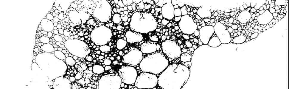
Rozpoznanie opiera się na wyniku badania histopatologicznego, poszerzonego o badania immunohistochemiczne, natomiast rozległość zmiany i jej lokalizację określa się po wykonaniu badań obrazowych. Nowotwór ten należy różnicować z innymi zmianami złośliwymi, głównie z naczyniakomięsakiem o znacznie gorszym rokowaniu niż RH. Guz wymaga różnicowania również ze zmianami zapalnymi, co może skutkować opóźnieniem wdrożenia prawidłowego leczenia.
Leczeniem z wyboru jest chirurgiczne usunięcie guza z ewentualną uzupełniającą radioterapią, bardzo rzadko z chemioradioterapią.
Celem pracy jest przedstawienie przypadku RH o bardzo rzadkim umiejscowieniu, jakim jest jama ustna, oraz zwrócenie uwagi na trudności diagnostyczne. Poniżej opisano przebieg kliniczny oraz proces diagnostyczno-leczniczy u 13-letniej pacjentki z naczyniakośródbłoniakiem siatkowatym szczęki prawej.
Hasła indeksowe: naczyniakośródbłoniak siatkowaty, nowotwór, leczenie chirurgiczne
Retiform hemangioendothelioma. Review of the literature and case study
Abstract
Retiform hemangioendothelioma (RH) is a rare vascular neoplasm of borderline malignancy, most commonly occurring in young adults. It frequently recurs locally, while distant metastases are rare. It is typically located within the skin and subcutaneous tissue of the lower limbs and only exceptionally affects the oral cavity.
The diagnosis is based on histopathological examination, supplemented by immunohistochemical testing, whereas the extent and location of the lesion are determined through imaging studies. This tumor must be differentiated from other malignant lesions, primarily angiosarcoma, which has a significantly
worse prognosis than RH. The tumor also requires differentiation from inflammatory lesions, which may result in delayed implementation of proper treatment.
The treatment of choice is surgical excision of the tumor, potentially with adjuvant radiotherapy, and very rarely with chemoradiotherapy.
The aim of this study is to present a case of RH in a very rare location—the oral cavity—and to highlight diagnostic difficulties. Below, we describe the clinical course and the diagnostic-therapeutic process in a 13-year-old female patient with retiform hemangioendothelioma of the right maxilla.
Key words: retiform hemangioendothelioma, neoplasm, surgical treatment
Piśmiennictwo:
- Fletcher CDM, Bridge JA, Hogendoorn PCW i wsp. (eds). WHO classification of tumours of soft tissue and bone. 5th ed. Lyon: IARC; 2020.
- Nobeyama Y, Ishiuji Y, Nakagawa H. Retiform hemangioendothelioma treated with conservative therapy: report of a case and review of the literature. Int J Dermatol. 2016; 55(2): 238-243.
- Calonje E, Fletcher CD, Wilson-Jones E i wsp. Retiform hemangioendothelioma: a distinctive form of low-grade angiosarcoma delineated in series of 15 cases. Am J Surg Pathol. 1994; 18(2): 115-125.
- Liu Q, Ouyang R, Chen P i wsp. A case report of retiform hemangioendothelioma as pleural nodules with literature review. Diagn Pathol. 2015; 10: 194.
- Tan D, Kraybill W, Cheney RT i wsp. Retiform hemangioendothelioma: a case report and review of the literature. J Cutan Pathol. 2005; 32(9): 634-637.
- Mondal A, Das M, Chatterjee U i wsp. Retiform hemangioendothelioma: an uncommon vascular neoplasm. Indian J Pathol Microbiol. 2020; 63(1): 122-124.
- Chen CW, Hsiao KH, Lu TJ i wsp. Retiform and epithelioid hemangioendothelioma arising from the spleen. Formos J Surg. 2019; 52(4): 147-150.
- Keiler SA, Honda K, Bordeaux JS. Retiform hemangioendothelioma treated with Mohs micrographic surgery. J Am Acad Dermatol. 2011; 65(1): 233-235.
- Hirsh AZ, Yan W, Wei L i wsp. Unresectable retiform hemangioendothelioma treated with external beam radiation therapy and chemotherapy: a case report and review of the literature. Sarcoma. 2010; 2010: 756246.
- Khade S, Rao M, Vishnoi JR i wsp. Retiform hemangioendothelioma: a rare tumor in the medial canthus: case report and review of literature. J Cutan Aesthet Surg. 2022; 15(3): 319-322.
- Emberger M, Laimer M, Steiner H i wsp. Retiform hemangioendothelioma: presentation of a case expressing D2-40. J Cutan Pathol. 2009; 36(9): 987-990.
- Parsons A, Sheehan DJ, Sangueza OP. Retiform hemangioendotheliomas usually do not express D2-40 and VEGFR-3. Am J Dermatopathol. 2008; 30(1): 31-33.
- Fanburg-Smith JC, Michal M, Partanen TA i wsp. Papillary intralymphatic angioendothelioma (PILA): a report of twelve cases of a distinctive vascular tumor with phenotypic features of lymphatic vessels. Am J Surg Pathol. 1999; 23(9): 1004-1010.
- Yarmel D, Dormans JP, Pawel BR i wsp. Recurrent pedal hobnail (Dabska-retiform) hemangioendothelioma with forefoot reconstructive surgery using a digital fillet flap. J Foot Ankle Surg. 2008; 47(5): 487-493.


