

Dostęp do tego artykułu jest płatny.
Zapraszamy do zakupu!
Po dokonaniu zakupu artykuł w postaci pliku PDF prześlemy bezpośrednio pod twój adres e-mail.
REPORTAŻ KLINICZNY
Leczenie chirurgiczne kostniaka żuchwy z wykorzystaniem dwufazowego siarczanu wapnia – opis przypadku
Surgical treatment of osteoid osteoma of the mandible using biphasic calcium sulfate – a case report
Damian Dudek, Maciej Jagielak, Aldona Chloupek, Oliwia Warmusz, Małgorzata Czarnecka, Edyta Reichman-Warmusz
Streszczenie
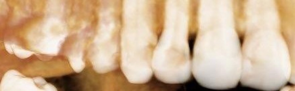
Znaczny odsetek patologii kości szczękowych wymaga leczenia chirurgicznego ze względu na rozpoznanie torbieli zębopochodnych lub guzów łagodnych. W grupie tej pewien procent mogą stanowić kostniaki. Są one częstymi, wolnorosnącymi zmianami patologicznymi, które z reguły są lepiej lub gorzej odgraniczone od otaczającej je kości. W zaprezentowanej pracy po usunięciu niewielkiego guza w połączeniu z resekcją wierzchołka korzenia zęba przedtrzonowego i wypełnieniem wstecznym kanału korzenia zęba znajdującego się w świetle zmiany umieszczono w ubytku twardniejący cement regeneracyjny w ramach sterowanej regeneracji tkanek, w celu przyspieszenia wzrostu nowej kości. Autorzy uważają zastosowanie takiego właśnie jednoetapowego leczenia kostniaka bocznego odcinka żuchwy jako optymalny w kontekście radykalności operacji oraz regeneracji ubytku kostnego związanego z powstałą zmianą chorobową.
Abstract
A significant percentage of jaw bone pathologies require surgical treatment due to the diagnosis of odontogenic cysts and benign tumors. A certain percentage of this group may consist of osteomas. They are common, slowly growing lesions that are usually better or worse demarcated from the surrounding bone. In the presented work, after removal of the small tumor in combination with apicoectomy in premolar tooth and retrograde filling of the tooth root canal located in the site of the lesion, hardening regenerative cement was placed in the defect as a guided tissue regeneration procedure to accelerate the growth of new bone. The authors consider the use of such a one-stage treatment of osteoma in the lateral part of the mandible as optimal in the context of the radical nature of the operation and the regeneration of the bone defect associated with the resulting lesion.
Hasła indeksowe: kostniak kostnawy, mikrochirurgia endodontyczna, dwufazowy siarczan wapnia
Key words: osteoid osteoma, endodontic micro surgery, biphasic calcium sulfate
Piśmiennictwo
1. Johnson NR, Gannon OM, Savage NW i wsp. Frequency of odontogenic cysts and tumors: a systematic review. J Investig Clin Dent. 2014; 5(1): 9-14.
2. Franklin JRB, Vieira EL, Brito LNS i wsp. Epidemiological evaluation of jaw cysts according to the new WHO classification: a 30-year retrospec tive analysis. Braz Oral Res. 2021; 35: e129.
3. Koenig LJ, Tamimi DF, Petrikowski CG i wsp. Diagnostic imaging: oral and maxillofacial. Wyd. 2. Philadelphia: PA: Elsevier; 2017.
4. Su YK, Wang J, Zhang TF i wsp. Odontogenic tumors and odontoge nic cysts: a clinical and pathological analysis of 4 181 cases. Zhong hua Kou Qiang Yi Xue Za Zhi. 2019;54(8):546-552.
5. Cheng C, Takahashi H, Yao K i wsp. Cemento-ossifying fibroma of maxillary and sphenoid sinuses: case report and literature review. Acta Oto laryngol Suppl. 2002;(547):118-22.
6. Waldron CA. Fibro-osseous lesions of the jaws. J Oral Maxillofac Surg. 1993;51(8):828-835.
7. Chang CC, Hung HY, Chang JY i wsp. Central ossifying fibroma: a clinicopathologic study of 28 cases. J Formos Med Assoc. 2008;107(4):288-94.
8. Vegas Bustamante E, Gargallo Albiol J, Berini Aytés L i wsp. Benign fibro-osseous lesions of the maxillas: analysis of 11 cases. Med Oral Pa tol Oral Cir Bucal. 2008;13(10):E653-6.
9. MacDonald-Jankowski DS. Fibro-osseous lesions of the face and jaws. Clin Radiol. 2004;59(1):11-25.
10. Nelson BL, Phillips BJ. Benign Fibro-Osseous Lesions of the Head and Neck. Head Neck Pathol. 2019;13(3):466-475.
11. Yahav A, Kurtzman GM, Katzap M, Dudek D iwsp. Bone Regeneration. Properties and Clinical Applications of Biphasic Calcium Sulfate. Dent Clin North Am. 2020; 64:453-472.
12. Dudek D, Reichman-Warmusz E, Kurtzman GM iwsp. The use of grafting material biphasic calcium sulfate for the treatment of osseous defects resulting from radicular cysts. Clinical study and six-month follow up. J Osteointegration. 2020; 12(4):716-721.
13. Azim AA, Albanyan H, Azim KA iwsp. The Buffalo study: Outcome and associated predictors in endodontic microsurgery – acohort study. Int Endod J. 2021;54(3):301-318.
14. DreesmanH.About bone healing [in German]. Beitr Klin Chir. 1892; 9: 804.
15. Baranes D, Kurtzman GM. Biphasic Calcium Sulfate as an Alternati ve Grafting Material in Various Dental Applications. J Oral Implantol. 2019;45(3):247-255.
16. Laino L, Troiano G, Giannatempo G iwsp. Sinus Lift Augmentation by Using Calcium Sulphate. ARetrospective 12 Months Radiographic Eva luation Over 25 Treated Italian Patients. Open Dent J. 2015; 9:414-419.
17. Stein JM, Fickl S, Yekta SS iwsp. Clinical evaluation of abiphasic cal cium composite grafting material in the treatment of human periodon tal intrabony defects. A12-month randomized controlled clinical trial. J Periodontol. 2009; 80(11):1774-1782.
18. Lombardo G, Marincola M, Cicconetti Aiwsp. Successful management of peri-implantitis around short and ultrashort single-crown implants: Acase series with a3-year follow-up. Int J Dent. 2019; 2019:5302752.
19. Mistry S, Roy S, Maitra NJ iwsp. Anovel, multi-barrier, drug eluting calcium sulfate/biphasic calcium phosphate biodegradable composite bone cement for treatment of experimental MRSA osteomyelitis in rab bit model. J Control Release. 2016; 239:169-181.


