

Dostęp do tego artykułu jest płatny.
Zapraszamy do zakupu!
Po dokonaniu zakupu artykuł w postaci pliku PDF prześlemy bezpośrednio pod twój adres e-mail.
STUDIA PRZYPADKÓW
Zębiak zestawny – aktualny problem kliniczny
Compound odontoma – current clinical problem
Joanna Trzcinka-Guźlińska, Łukasz Woźniak, Milena Zabłocka, Jan Borys, Bożena Antonowicz
Streszczenie
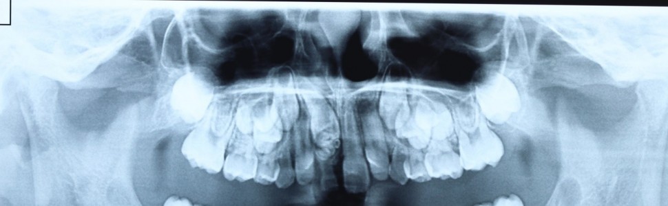
Zębiaki to jedne z najczęstszych guzów zębopochodnych występujących w jamie ustnej. Ich etiologia nie jest do końca poznana. Większość autorów dzieli je na zębiaki złożone i zestawne. Najczęstszym objawem sugerującym występowanie zębiaka jest zaburzone wyrzynanie zębów stałych. Wykrywane są zazwyczaj przypadkowo na zdjęciach rentgenowskich. W artykule został przedstawiony przypadek 7-letniego pacjenta, który zgłosił się z rodzicem do poradni w celu konsultacji i leczenia zmiany, wykrytej na zdjęciu rentgenowskim, nad przetrwałym zębem mlecznym 51. Po analizie badania podmiotowego, przedmiotowego oraz ocenie zdjęcia pantomograficznego zaplanowano zabieg wyłuszczenia guza, wykonanie badania histopatologicznego oraz usunięcie przetrwałego zęba mlecznego 51. Zatrzymany ząb 11 pozostawiono do obserwacji i samoistnej erupcji. W pracy autorzy zwracają uwagę na fakt występowania w praktyce klinicznej zębiaków i konieczność prawidłowej diagnostyki i leczenia tej anomalii. W celu prawidłowego rozpoznania i leczenia zębiaków konieczna jest współpraca doświadczonego specjalisty chirurgii stomatologicznej lub szczękowo-twarzowej oraz lekarza ortodonty.
Abstract
Odontomas are among the most common odontogenic tumors found in the oral cavity. Their etiology is not fully understood. Majority of authors divide them into complex and compound odontomas The most frequent symptom suggesting the occurrence of an odontoma is the disturbance in permanent tooth eruption. They are typically detected accidentally on radiographic images. The paper presents the case of a 7-year-old patient who visited the clinic for consultation and treatment of a lesion detected on an X-ray over a persistent deciduous tooth 51. After the subjective and physical examination, pantomographic image analysis, the procedure of enucleation of the tumor and histopathological examination as well as the extraction of the persistent tooth 51 were made. The retained tooth 11 was left for observation and autonomous eruption. Authors of the paper point to the fact of the presence of odontomas in the clinical practice and the need for the correct diagnostics and treatment of this anomaly. In order to correctly diagnose and treat odontomas, the cooperation of an experienced specialist in dental surgery and an orthodontist is necessary.
Hasła indeksowe: zębiak zestawny, chirurgia stomatologiczna, anomalie zębowe, opis przypadku, guz zębopochodny
Key words: compound odontoma, dental surgery, dental anomalies, case report, odontogenic tumor
Piśmiennictwo
- Kaczmarzyk T, Stypułkowska J, Tomaszewska R i wsp. Nowotwory zębopochodne i guzy nowotworopodobne kości szczękowych. Warszawa: Kwintesencja;
- da Silva VA, Pedreira RP, Sperandio FF i wsp. Odontomas are associated with impacted permanent teeth in orthodontic patients. J Clin Exp Dent. 2019; 11(9): e790-e794.
- Kämmerer PW, Schneider S, Schiegnitz E i wsp. Clinical parameter of odontoma with special emphasis on treatment of impacted teeth – a retrospective multicentre study and literature review. Clin Oral Investig. 2016; 20(7): 1827-1835.
- Gupta A, Vij H, Vij R i wsp. An erupted compound odontoma. BMJ Case Rep. 2014; 2014: bcr2013201820.
- Karolak D, Lange J. Leczenie interdyscyplinarne (chirurgiczno-ortodontyczne) zębiaka zestawnego związanego z zatrzymanym stałym siekaczem bocznym żuchwy – opis przypadku. Nowa Stomatol. 2018; 23(1): 25-31.
- Katz RW. An analysis of compound and complex odontomas. ASDC J Dent Child. 1989; 56(6): 445-449.
- Nelson BL, Thompson LD. Compound odontoma. Head Neck Pathol. 2010; 4(4): 290-291.
- Soluk Tekkesin M, Pehlivan S, Olgac V i wsp. Clinical and histopathological investigation of odontomas. Review of the literature and presentation of 160 cases. J Oral Maxillofac Surg. 2012; 70(6): 1358-1361.
- Zaleska M, Gronkiewicz K, Blat J i wsp. Skojarzone leczenie zębiaka szczęki – opis przypadku. Endodoncja w Praktyce. 2017; 1: 48-55.
- Machado Cde V, Knop LA, da Rocha Mc i wsp. Impacted permanent incisors associated with compound odontoma. BMJ Case Rep. 2015; 2015: bcr2014208201.
- Gururaju CR, Rathva Vj, Usha C i wsp. A compound odontoma in the path of an erupting incisor. BMJ Case Rep. 2013; 2013: bcr2013200825.
- Tyagi P, Singla S. Complex composite odontoma. Int J Clin Pediatr Dent. 2010; 3(2): 117-120.
- Amado Cuesta S, Gargallo Albiol J, Berini Aytés L i wsp. Review of 61 cases of odontoma. Presentation of an erupted complex odontoma. Med Oral. 2003; 8(5): 366-373.
- Serra-Serra G, Berini-Aytés L, Gay-Escoda C. Erupted odontomas. A report of three cases and review of the literature. Med Oral Patol Oral Cir Bucal. 2009; 14(6): E299-E303.
- Bereket C, Çakır-Özkan N, Şener İ i wsp. Complex and compound odontomas. Analysis of 69 cases and a rare case of erupted compound odontoma. Niger J Clin Prac 2015; 18(6): 726-730.
- Preoteasa CT, Preoteasa E. Compound odontoma – morphology, clinical findings and treatment. Case report. Rom J Morphol Embryo 2018; 59(3): 997-1000.
- Sreedharan S, Krishnan IS. Compound odontoma associated with impacted maxillary incisors. J Indian Soc Pedod Prev Dent. 2012; 30(3): 275-278.
- Jayam C, Bandlapalli A, Patel N i wsp. A case of impacted central incisor due to dentigerous cyst associated with impacted compound odontome. BMJ Case Rep. 2014; 2014: bcr2013202447.
- Janas A, Grzesiak-Janas G. Zębiaki złożone. Dent Med Probl. 2005; 42(3): 425-429.
- Reichart PA, Philpsen HP. Odontogenic tumors and allied lesions. Batavia, IL: Quintessence Publishing; 2004.


