

Dostęp do tego artykułu jest płatny.
Zapraszamy do zakupu!
Po dokonaniu zakupu artykuł w postaci pliku PDF prześlemy bezpośrednio pod twój adres e-mail.
Rehabilitacja protetyczna u pacjentów z nabytym ubytkiem tkanek kości szczęk. Opis przypadków
Prosthetic rehabilitation of patients with acquired maxillary defect – case reports
Józef Listwan, Andrzej Gala
Streszczenie
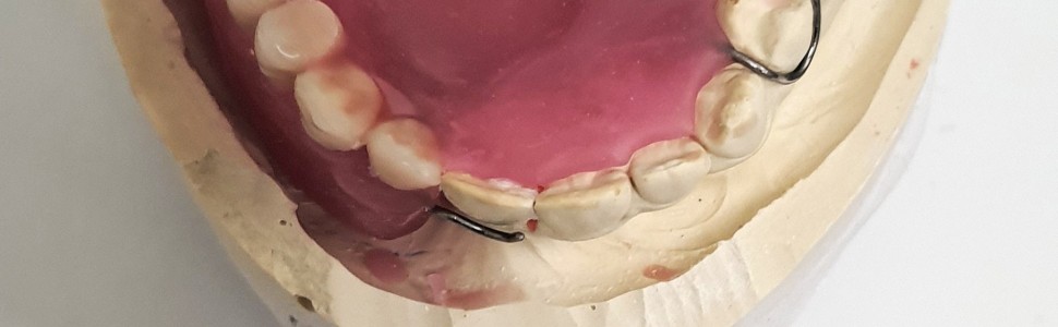
Protetyczna rehabilitacja ubytków poresekcyjnych kości szczęk stanowi wyzwanie ze względu na utratę tkanek podłoża protetycznego, uniemożliwiającą uzyskanie odpowiedniej stabilizacji oraz retencji, jak w przypadku konwencjonalnych protez. Leczenie chirurgiczne nie zawsze jest możliwe, z tego powodu wciąż wielu pacjentów wymaga leczenia z zastosowaniem obturatorów protetycznych. W poniższej pracy omówione zostały ogólne zasady postępowania w trakcie rehabilitacji pacjentów poresekcyjnych, opisano wybrane aspekty konstrukcji aparatów protetycznych oraz ograniczenia związane z przylegającymi strukturami anatomicznymi. W artykule przedstawiono również dwa przypadki leczenia pacjentów z brakami w obrębie kości środkowego piętra twarzy oraz obecnym połączeniem ustno-nosowym powstałym po zabiegu operacyjnym. Głównym celem zastosowanej terapii była możliwość przywrócenia fizjologicznych funkcji, w szczególności odtworzenie funkcji mowy, żucia oraz połykania. Nie bez znaczenia pozostaje również aspekt estetyczny oraz psychologiczny przekładający się na jakość życia i samopoczucie pacjenta. Terapia protetyczna w tej szczególnej grupie pacjentów często wymaga rozwiązań niekonwencjonalnych, dostosowanych do indywidualnych warunków pacjenta. Powodzenie leczenia zależy także od dobrej komunikacji oraz współpracy z innymi specjalistami, takimi jak chirurg szczękowo-twarzowy, logopeda, fizjoterapeuta i psycholog.
Abstract
Prosthetic rehabilitation of post-resection defects in the jaw bones is a challenge due to the tissue loss of denture space that does not allow for adequate stabilization and retention as with conventional dentures. Surgical treatment is not always possible, therefore many patients still require treatment with prosthetic obturators. The following article discusses the general principles of proceeding during the rehabilitation of post-resection patients, brings up the selected aspects of the construction of prosthetic appliances and the limitations associated with adjacent anatomical structures. The article also presents two cases of the treatment of patients with missing bones in the middle level of the face and the present oronasal connection after maxillary surgery. The main goals of described therapy were the possibility of restoring physiological functions, in particular the functions of speech, chewing and swallowing. The aesthetic and psychological aspects of the patient, which translates into the quality of life and well-being, is also important. Prosthetic therapy in this special group of patients often requires unconventional solutions adapted to the individual conditions of the patient. The success of treatment also depends on good communication and cooperation with other specialists, such as the maxillofacial surgeon, speech therapist, physiotherapist and psychologist.
Hasła indeksowe: rehabilitacja protetyczna, obturator protetyczny, resekcja szczęki
Key words: prosthetic rehabilitation, prosthetic obturator, maxilla resection
Piśmiennictwo
- Rolski D, Juszczyszyn K, Nieborak R i wsp. Rehabilitacja implantoprotetyczna pacjentów po operacjach nowotworów w obrębie głowy i szyi – obserwacje odległe. Protet Stomatol. 2017; 67(3): 255-269.
- Maślak-Bereś M, Loster JE. Clinical protocol during prosthetic treatment of patients with tissue deficiences in oral and facial areas. Prosthodontics. 2019; 69(3): 322-331.
- Siddall KZ, Rogers SN, Butterworth CJ. The prosthodontic pathway of the oral cancer patient. Dent Update. 2012; 39(2): 98-100.
- Juszczyszyn K, Rolski D, Mierzwińska-Nastalska E. The use of CAD/CAM technology in prosthetic rehabilitation of patients treated for maxillary tumors. Prosthodontics. 2019; 69(3): 313-321.
- Brandão TB, Vechiato Filho AJ, Batista VE i wsp. Obturator prostheses versus free tissue transfers. A systematic review of the optimal approach to improving the quality of life for patients with maxillary defects. J Prosthet Dent. 2016; 115(2): 247-253.
- Depprich R, Naujoks C, Lind D i wsp. Evaluation of the quality of life of patients with maxillofacial defects after prosthodontic therapy with obturator prostheses. Int J Oral Maxillofac Surg. 2011; 40(1): 71-79.
- Ali A, Fardy MJ, Patton DW. Maxillectomy – to reconstruct or obturate? Results of a UK survey of oral and maxillofacial surgeons. Br J Oral Maxillofac Surg. 1995; 33(4): 207-210.
- Okay DJ, Genden E, Buchbinder D i wsp. Prosthodontic guidelines for surgical reconstruction of the maxilla. A classification system of defects. J Prosthet Dent. 2001; 86(4): 352-363.
- Dos Santos DM, de Caxias FP, Bitencourt SB i wsp. Oral rehabilitation of patients after maxillectomy. A systematic review. Br J Oral Maxillofac Surg. 2018; 56(4): 256-266.
- Jung S, Kook MS, Park HJ i wsp. Delayed closure of the palatal defect using buccal inversion and palatal rotation flaps after maxillectomy. J Craniofac Surg. 2013; 24(2): 660-664.
- Keyf F. Obturator prostheses for hemimaxillectomy patients. J Oral Rehabil. 2001; 28(9): 821-829.
- Ali R, Altaie A, Nattress B. Rehabilitation of oncology patients with hard palate defects, part 3: Construction of an acrylic hollow box obturator. Dent Update. 2015; 42(7): 612-620.
- Sharma AB, Beumer J 3rd. Reconstruction of maxillary defects. The case for prosthetic rehabilitation. J Oral Maxillofac Surg. 2005; 63(12): 1770-1773.
- Spiechowicz E. Protetyka Stomatologiczna. Warszawa: Wydawnictwo Lekarskie PZWL; 2008: 488-500.
- Kornblith AB, Zlotolow IM, Gooen J i wsp. Quality of life of maxillectomy patients using an obturator prosthesis. Head Neck. 1996; 18(4): 323-334.
- Parr GR, Tharp GE, Rahn AO. Prosthodontic principles in the framework design of maxillary obturator prostheses. J Prosthet Dent. 2005; 93(5): 405-411.
- Gay WD, King GE. Applying basic prosthodontic principles in the dentulous maxillectomy patient. J Prosthet Dent. 1980; 43(4): 433-435.
- Desjardins RP. Obturator prosthesis design for acquired maxillary defects. J Prosthet Dent. 1978; 39(4): 424-435.
- Devlin H, Barker GR. Prosthetic rehabilitation of the edentulous patient requiring a partial maxillectomy. J Prosthet Dent. 1992; 67(2): 223-227.
- Kucharski Z, Rolski D. Zastosowanie kliniczne materiałów elastycznych do podścieleń ruchomych uzupełnień protetycznych. Protet Stomatol. 2011; 61(3): 234-240.
- Djan R, Penington A. A systematic review of questionnaires to measure the impact of appearance on quality of life for head and neck cancer patients. J Plast Reconstr Aesthet Surg. 2013; 66(5): 647-659.
- Rogers SN, Ahad SA, Murphy AP. A structured review and theme analysis of papers published on ‘quality of life’ in head and neck cancer: 2000-2005. Oral Oncol. 2007; 43(9): 843-868.
- Wang RR. Sectional prosthesis for total maxillectomy patients. A clinical report. J Prosthet Dent. 1997; 78(3): 241-244.
- Artopoulou II, Karademas EC, Papadogeorgakis N i wsp. Effects of sociodemographic, treatment variables, and medical characteristics on quality of life of patients with maxillectomy restored with obturator prostheses. J Prosthet Dent. 2017; 118(6): 783-789.
- Rogers SN, Lowe D, McNally D i wsp. Health-related quality of life after maxillectomy. A comparison between prosthetic obturation and free flap. J Oral Maxillofac Surg. 2003; 61(2): 174-181.
- Eckardt A, Teltzrow T, Schulze A i wsp. Nasalance in patients with maxillary defects – reconstruction versus obturation. J Craniomaxillofac Surg. 2007; 35(4-5): 241-245.
- Moreno PI, Ramirez AG, San Miguel-Majors SL i wsp. Satisfaction with cancer care, self-efficacy, and health-related quality of life in Latino cancer survivors. Cancer. 2018; 124(8): 1770-1779.
- Moreno MA, Skoracki RJ, Hanna EY i wsp. Microvascular free flap reconstruction versus palatal obturation for maxillectomy defects. Head Neck. 2010; 32(7): 860-868.
- Wu YQ, Huang W, Zhang ZY i wsp. Clinical outcome of dental implants placed in fibula-free flaps for orofacial reconstruction. Chin Med J (Engl). 2008; 121(19): 1861-1865.
- Wang F, Huang W, Zhang C i wsp. Functional outcome and quality of life after a maxillectomy. A comparison between an implant supported obturator and implant supported fixed prostheses in a free vascularized flap. Clin Oral Implants Res. 2017; 28(2): 137-143.
- Nyberg EL, Farris AL, Hung BP i wsp. 3D-printing technologies for craniofacial rehabilitation, reconstruction, and regeneration. Ann Biomed Eng. 2017; 45(1): 45-57.
- Kortes J, Dehnad H, Kotte ANT i wsp. A novel digital workflow to manufacture personalized three-dimensional-printed hollow surgical obturators after maxillectomy. Int J Oral Maxillofac Surg. 2018; 47(9): 1214-1218.
- Rodney J, Chicchon I. Digital design and fabrication of surgical obturators based only on preoperative computed tomography data. Int J Prosthodont. 2017; 30(2): 111-112.
- Elbashti ME, Hattori M, Patzelt SB i wsp. Feasibility and accuracy of digitizing edentulous maxillectomy defects. A comparative study. Int J Prosthodont. 2017; 30(2): 147-149.
- Al Mardini M. Prosthetic rehabilitation of the head and neck. The state of the art. Curr Opin Otolaryngol Head Neck Surg. 2009; 17(4): 253-257.


