

Dostęp do tego artykułu jest płatny.
Zapraszamy do zakupu!
Po dokonaniu zakupu artykuł w postaci pliku PDF prześlemy bezpośrednio pod twój adres e-mail.
Aneta Olszewska, Damian Korczyński
Streszczenie
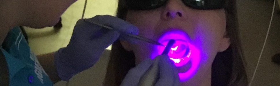
Lasery diodowe znane są z szerokiego zastosowania w leczeniu chirurgicznym, periodontologicznym i endodontycznym u dorosłych pacjentów. Obecnie, ze względu na obecność na rynku laserów o różnej długości fali, możliwości terapeutyczne laserów diodowych pozwalają na coraz szersze zastosowanie również u młodych pacjentów. Laseroterapia stwarza bardziej komfortowe i mniej stresujące warunki pracy dla lekarza i dziecka, takie jak: zmniejszenie dolegliwości bólowych, łatwa i szybka aplikacja, efekt hemostatyczny, mniejsze dolegliwości pozabiegowe, szybsze gojenie i ograniczona potrzeba stosowania antybiotyków. W pracy przedstawiono wykorzystanie lasera diodowego w różnych procedurach stomatologicznych u dzieci.
Abstract
Diode lasers are very well known for their wide range of applications in surgery, periodontics and endodontics for adults. Nowadays, diode laser has one of the most versatile ranges of wavelengths available due to the number of different therapies that can be performed for children and adolescents. . They provide a comfortable and stress-free environment to the pedodontist and a child by offering reduced pain, swelling, easy application ,faster healing, an excellent haemostatic effect and the reduced use of post-operative antibiotics. The aim of this paper is to review the various wavelengths diode laser applications in pediatric dentistry.
Hasła indeksowe: laser diodowy, stomatologia dziecięca, długość fali promieniowania
Key words: diode laser, pediatric dentistry, laser wavelengths
PIŚMIENNICTWO
- AAPD: Policy on the Use of Lasers for Pediatric Dental Patients. Review Council Council on Clinical Affairs. Latest Revision 2017. The Reference Manual of Pediatric Dentistry. 2020-2021: 116-118.
- Galui S, Pal S, Mahata S i wsp. Laser and its use in pediatric dentistry: A review of literature and a recent update. Int J Pedod Rehabil. 2019; 4: 1-5.
- Olivi G, Margolis FS, Genovese MD. Pediatric Laser Dentistry; A User’s Guide. Chicago: Quintessence Publishing; 2011: 73-76.
- Norbert G, Rene F, Leon V i wsp. Laser in pediatric dentistry – a review. J Oral Laser Appl. 2005; 5: 207-209.
- Koenig K, Schneckenburger H. Laser-induced autofluorescence for medical diagnosis. J Fluoresc. 1994; 4(1): 17-40. doi: 10.1007/BF01876650
- Volgenant CM, van der Veen MH, de Soet JJ i wsp. Effect of metalloporphyrins on red autofluorescence from oral bacteria. Eur J Oral Sci. 2013; 121(3 Pt 1): 156-161. doi: 10.1111/eos.12045
- Lennon AM, Buchalla W, Brune L i wsp. The ability of selected oral microorganisms to emit red fluorescence. Caries Res. 2006; 40(1): 2-5. doi: 10.1159/000088898
- Buchalla W, Attin T, Niedemann Y i wsp. Porphyrins are the cause of red fluorescence of carious dentine. Caries Res. 2008; 42: 223.
- König K, Flemming G, Hibst R. Laser-induced autofluorescence spectroscopy of dental caries. Cellular and Molecular Biology. (Noisy-le-Grand, France) 1998; 44(8): 1293-1300.
- Buchalla W, Lennon AM, Attin T. Comparative fluorescence spectroscopy of root caries lesions. Eur J Oral Sci. 2004; 112(6): 490-496.
- Buchalla W. Comparative fluorescence spectroscopy shows differences in non-cavitated enamel lesions. Caries Res. 2005; 39(2): 150-156.
- Braga M, Nicolau J, Rodriguez CR i wsp. Laser fluorescence devise does not perform well in detection of early caries lesions in primary teeth: an in vitro study. Oral Health Prev Dent. 2008; 6: 165-169.
- Ando M, van Der Veen MH, Schemehron BR i wsp. Comparative study to quantify demineralized enamel in deciduous and permanent teeth using laser and light-induced fluorescence techniques. Caries Res. 2001; 35: 464-470.
- Volgenant CM, van der Veen MH, de Soet JJ i wsp. Effect of metalloporphyrins on red autofluorescence from oral bacteria. Eur J Oral Sci. 2013; 121(3 Pt 1): 156-161. doi: 10.1111/eos.12045
- Lennon AM, Buchalla W, Brune L i wsp. The ability of selected oral microorganisms to emit red fluorescence. Caries Res. 2006; 40(1): 2-5. doi: 10.1159/000088898
- Zacharia MA, Munshi AK. Microbiological assessment of dentin stained with a caries detector dye. J Clin Pediatr Dent. 1995; 19: 111-115.
- Lennon AM, Buchalla W, Rassner B i wsp. Efficiency of 4 caries excavation methods compared. Oper Dent. 2006; 31(5): 551-555. doi: 10.2341/05-92
- Lennon AM, Attin T, Buchalla W. Quantity of remaining bacteria and cavity size after excavation with FACE, caries detector dye and conventional excavation in vitro. Oper Dent. 2007; 32(3): 236-241. doi: 10.2341/06-64
- Lennon AM, Attin T, Martens S i wsp. Fluorescence-aided caries excavation (FACE), caries detector, and conventional caries excavation in primary teeth. Pediatr Dent. 2009; 31(4): 316-319.
- Gimenez T, Braga MM, Raggio DP i wsp. Fluorescence based methods for detecting caries lesions: Systematic review, meta-analysis and sources of heterogeneity. PLoS One. 2013; 8(4,4): e60421. doi: 10.1371/journal.pone.0060421
- Moritz A. Oral Laser Application. Quintessence; 2006: 528-529.
- Boj J, Galofre N, Espana A i wsp. Pain perception in paediatric patients undergoing laser treatments. J Oral Laser Appl. 2005; 2: 85-89.
- Koci E, Almas A. Laser application in dentistry: an evidence-based clinical decision-making update. Pak Oral Dent J. 2009; 29(2): 409-423.
- Olivi G, Genovese MD, Caprioglio C. Evidence-based dentistry on laser paediatric dentistry: review and outlook. Eur J Paediatr Dent. 2009; 10(1): 29-40. doi: 10.1007/bf03262783
- Boj J. The Future Of Laser Pediatric Dentistry. J Oral Laser Appl. 2005; 5: 173-7.
- Koci E, Almas A. Laser application in dentistry: an evidence-based clinical decision-making update. Pak Oral Dent J. 2009; 29(2): 409-423.
- Kotlow L. Lasers in pediatric dentistry. General Dent. 2009; 140-146.
- Cruz DR, Kohara EK, Ribeiro MS i wsp. Effects of low intensity laser therapy on the orthodontic movement velocity of human teeth: a preliminary study. Lasers Surg Med. 2004; 35(2): 117-120. doi: 10.1002/lsm.20076
- O’Neill JF, Hope CK, Wilson M. Oral bacteria in multispecies biofilm can be killed by red light in the presence of toluidine blue. Lasers Surg Med. 2002; 31: 86-90.
- Bonsor SJ, Nichol R, Reid TM i wsp. Microbiological evaluation of photo-activated disinfection in endodontics (an in vivo study). Br Dent J. 2006; 200(6): 337-341.
- Odor TM, Chandler NP, Watson TF i wsp. Laser light transmission in teeth: a study of the patterns in different species. Int Endodontic Journal. 1999; 32: 296-302.
- Burns T, Wilson M, Pearson GJ. Effect of dentin and collagen on the lethal photosensitization of Streptococcus mutans. Caries Res. 1995; 29: 192-197.
- Soares F, Varella CH, Pileggi R i wsp. Impact of Er,Cr:YSGG laser therapy on the cleanliness of the root canal walls of primary teeth. J Endod. 2008; 34(4): 474-477. doi:10.1016/j. joen.2008.02.006
- Haas R, Baron M, Dortbudak O i wsp. Lethal photosensitisation, autologous bone and e-PTFE membrane for the treatment of peri-implantitis: preliminary results. Laryngoscope 2000; 105: 867-871.
- Ramazani N, Ahmadi R, Daryaeian M. Oral and dental laser treatment for children: applications, advantages and consideration. J Lasers Med Sci. 2012; 3(1): 44-49.
- Pakins F. Lasers in pediatric and adolescent dentistry. Dent Clin North Am. 2000; 44(4): 821-830.


