

Dostęp do tego artykułu jest płatny.
Zapraszamy do zakupu!
Po dokonaniu zakupu artykuł w postaci pliku PDF prześlemy bezpośrednio pod twój adres e-mail.
Streszczenie
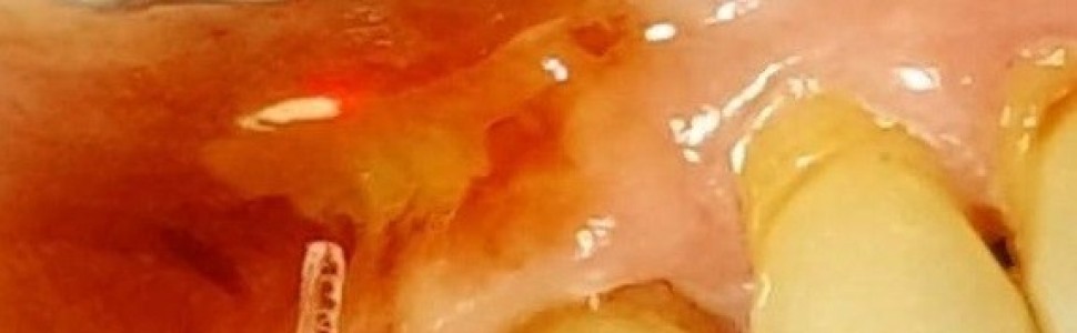
Celem niniejszej pracy jest prezentacja przypadku i ocena efektywności zastosowania lasera Er:YAG o długości fali 2940 nm (AdvErL EVO , Morita, Kioto, Japonia) w leczeniu postaci tarczkowatej liszaja płaskiego, obecnego na błonie śluzowej jamy ustnej. Do usunięcia zmiany użyto następujących parametrów fizycznych ustawień lasera: 20Hz, 80mJ, 1,6 W, impuls 300 µs, woda 8, powietrze 7 oraz końcówkę szklaną, płaską o średnicy 800µm. Osiągnięto znaczną redukcję wielkości patologicznejzmiany pracując techniką kontaktową, warstwa po warstwie, bez jakichkolwiek powikłań w okresie pozabiegowym. Zastosowanie tej długości fali zapewniło zabiegowi cechy minimalnie inwazyjnego. Przedstawiona metoda, jako leczenie uzupełniające w keratotycznych formach liszja płaskiego wydaje się być interesującą i skuteczną alternatywądla konwencjonalnego leczenia. Z racji jednak na niewielką ilość doniesień w dostępnym piśmiennicwie, zaproponowana technikawymaga przeprowadzenia większej ilości zabiegów i dłuższej obserwacji klinicznej.
Abstract
The aim of this study is to present a case and evaluate the effectiveness of the Er:YAG laser with a wavelength of 2940 nm (AdvErL EVO, Morita, Kyoto, Japan) in the treatment of the plaque Oral Lichen Planus (OLP) present on the oral mucosa. The following physical parameters of the laser settings were used to remove lesions: 20 Hz, 80 mJ, 1.6 W, pulse duration 300 µs, water 8, air 7, and a flat, glass tip with a diameter of 800 µm. A significant reduction in the size of the pathological lesion was achieved by using a contact technique, layer by layer, without any complications during the postoperative period. The use of this wavelength provided minimally invasive treatment. The presented method, as a complementary treatment in keratotic forms of OLP, seems to be an interesting and effective alternative to conventional treatment. However, due to the limited numbers of cases in the literature, the proposed technique requires more procedures and longer clinical observation.
Hasła indeksowe: liszaj płaski jamy ustnej, laser Er:YAG, laseroterapia.
Key words: oral lichen planus, Er:YAG laser, laser therapy.
PIŚMIENNICTWO
1. Cordova P, Rubio A, Echeverria P. Orallichenplanus: a look from diagnosis to treatment. J Oral Res. 2014; 3(1): 62-67.
2. Torti D, Jorizzo J, McCarthy M. Orallichenplanus: a caseseries with emphasis on therapy. Arch Dermatol. 2007; 143(4): 511-515.
3. Krasowska D, Pietrzak A, Surdacka A i wsp. Psychologicalstressendocrine and immuneresponse in patiens with lichenplanus.Int J Dermatol. 2008; 47(11): 1126-1134.
4. Pietkowicz B, Sidor K, Berger M. Rola stresu w etiologii liszaja płaskiego jamy ustnej. StomatolWspółcz. 2011; 18(5): 41-46.
5. Roopashree M, Gondhalekar R, Shashikanth M i wsp.Pathogenesis of orallichenplanus – a review. J OralPatholMed. 2010; 39(10): 729-734.
6. Andreasen J. Orallichenplanus. 1. A clinicalevaluation of 115 cases. OralSurgOral Med Oral Pathol. 1968; 25(1): 31-42.
7. Mollaoglu N.Orallichenplanus: a review. Br J OralMaxillofac Surg. 2000; 38(4):370-377.
8. Eisen D. The clinicalmanifestations and treatment oforallichenplanus. Dermatol Clin. 2003; 21(1): 89.
9. Anuradha Ch, Reddy B, Nandan S i wsp. Orallichenplanus. A review. NYStateDent J. 2008; 74(4): 66-68.
10. Araya M, RojasAlcayaga G, Esguep A. Associationbetweenpsychologicaldisorders and the presence of orallichenplanus, burningmouthsyndrome and recurrentaphtousstomatitis. Med Oral. 2004; 9(1): 1-7.
11.Carrozzo M, Porter S, Mercadante V i wsp.Orallichenplanus: A diseaseor a spectrum of tissuereactions?Types, causes, diagnostic, algorhythms, prognosis, management strategies. Periodontol 2000. 2019; 80(1): 105-125.
12. Fitzpatrick S, Hirsch S, Gordon S. The malignanttransformation of orallichenplanus and orallichenoidlesions. A systematicreview. J Am DentAssoc. 2014; 145(1): 45-56.
13. Sulewska M, Duraj E, Sobaniec S i wsp. Ocena własnego protokołu terapii fotodynamicznej w leczeniu liszaja płaskiego języka. Laser. 2019; 1: 28-35.
14. Zegarelli D. The treatment of orallichenplanus.Ann Dent. 1993; 52(2): 3-8.
15.Ishida C, Ramos–e–Silva M.Cryosurgeryinorallesions. Int J Dermatol. 1998;37(4): 283-285.
16. White JM, Chaudhry SI, Kudler i wsp.Nd:YAG and CO2 laser therapy of oralmucosallesions. JClin Laser MedSurg. 1998;16(6): 299-304.
17. Sobaniec S, Bednarczyk P, Pietruski J i wsp. Clinicalassessment of the efficacy of photodynamictherapy in the treatment of orallichenplanus. Lasers Med Sci. 2013; 28(1): 311-316.
18. Mostafa D, Moussa E, Alnouaem M. Evaluation of photodynamictherapy in treatment of oral erosivelichenplanus in comparison with topical applied corticosteroids. PhotodiagnosisPhotodynTher. 2017;19: 56-66.
19. Jajarm H, Falaki F, Sanathani i wsp. A comparativestudy of toluidyne bluemediatedphotodynamictherapy versus topicalcorticosteroids in the treatment of erosive-atrophicorallichenplanus: a randomizedclinicalcontrolledtrial. Lasers Med Sci. 2015;30(5): 1475-1480.
20. Passeron T, Zakaria W, Ostovari N i wsp.Treatment of erosiveorallichenplanus by the 308 nmexcimer laser. Laser Surg Med. 2004;34(3):205.
21. Othman N, Shaker O, Elshenawy H i wsp. The effect of diode laser and topical steroid on serum level of TNF-alpha in orallichenplanuspatients. J ClinExp Dent. 2016; 8(5):e566-e570.
22. Jajarm H, Falaki F, Mahdavi O. A comparative pilot study of lowintensity laser versus topicalcorticosteroids in the treatment of erosive-atrophicorallichenplanus. Photomed Laser Surg. 2011; 29(6): 421-425.
23. Dillenburg C, Martins M, Munerato M i wsp.Efficacy of laser phototherapy in comparison to topicalclobetasol for the treatment of orallichenplanus: a randomizedcontrolledtrial. J BiomedOpt. 2014; 19(6): 068002.
24. Kazancioglu H, Erisen M. Comparison of low-level laser therapy versus ozonetherapy in the treatment of orallichenplanus. Ann Dermatol. 2015; 27(5): 485-491.
25. Van der Hem P, Egges M, Van der Waal J iwsp. CO2 laser evaporation of orallichenplanus. Int J OralMaxillofac Surg. 2008;37(7): 630-633.
26. Fornaini C, Raybaud H, Augros C i wsp. New ClinicalApproach for Use of Er:YAG Laser in the SurgicalTreatment of OralLichenPlanus: A Report of TwoCases. Photomed Laser Surg.2012;30(4): 234-238.
27. Frame J, Das Gupta A, Dalton G i wsp.Use of the carbondioxide laser in themanagement of premalignantlesions of the oralmucosa. J.LaryngolOtol. 1984; 98(12): 1251-1260.
28. Chen P, Toroian D, McKittrick J. Minerals Form a Continuum Phase in MatureCancellousBone.CalcifTissueInt.2011; 88(5):351-361.
29. Walsh JT Jr, Flotte TJ, Deutsch TF. Er:YAG laser ablation of tissue: effect of pulseduration and tissuetype on thermaldamage. LasersSurg Med. 1989; 9(4): 314-326.
30. Ishikawa I, Aoki A, Takasaki A i wsp. Application of lasers in periodontics: trueinnovationormyth? Periodontol 2000. 2009;50(1): 90-126.


