Dostęp do tego artykułu jest płatny.
Zapraszamy do zakupu!
Po dokonaniu zakupu artykuł w postaci pliku PDF prześlemy bezpośrednio pod twój adres e-mail.
Streszczenie
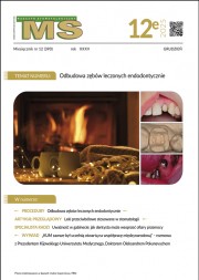
Wstęp. Istotnym elementem leczenia endodontycznego, oprócz opracowania i skutecznej dezynfekcji, jest szczelna obturacja kanałów korzeniowych. Ogromny postęp, jaki dokonuje się obecnie w endodoncji, wiąże się z systematycznym poszerzaniem asortymentu środków dostępnych na rynku stomatologicznym. Ciągłe pojawianie się nowych preparatów skłoniło autorów do usystematyzowania aktualnej wiedzy o materiałach do ostatecznego wypełniania kanałów korzeniowych.
Materiały i metody. Przeprowadzono analizę dostępnego piśmiennictwa, wyszukując hasła związane z materiałami do ostatecznego wypełniania kanałów korzeniowych w bazach Medline (PubMed) oraz ISI Web of Science.
Wnioski. W dalszym ciągu brakuje materiału, który stanowiłby – jako jedyny – wypełnienie systemu kanałów korzeniowych. Powszechnie zaleca się stosowanie jednocześnie dwóch materiałów: podstawowego – gutaperki oraz dodatkowego – pasty uszczelniającej przestrzenie między materiałem głównym a zębiną korzeniową.
Abstract
An important part of endodontic treatment, besides preparation and effective disinfection is solid obturation of the root canal system. Great progress is currently engaged in endodontics, steadily increasing range of resources available on the dental market. Constantly emerging new products prompted the authors to systematize the currently available knowledge about materials used for the final root canals obturation.
Materials and methods. A comprehensive search of Medline (PubMed) and ISI Web of Science databases was carried out with terms according to root canal filling materials
Conclusions. There is still no material that would serve as the sole filling of the root canal system. Generally it is recommended to use simultaneously two materials – base – gutta-percha and an additional – paste, sealing the spaces between the main material and the root dentine.
Hasła indeksowe: wypełnienie kanału korzeniowego, materiały biozgodne, materiały do ostatecznego wypełnienia kanałów korzeniowych
Key words: root canal obturation, biocompatible materials, root canal filling materials
PIŚMIENNICTWO
- Holland R. i wsp.: Factors affecting the periapical healing process of endodontically treated teeth. J. Appli. Oral Sci.: Revista FOB, 2017, 25, 465-476.
- Grossman L.: Endodontic failures. Dental Clin. North Am., 1972, 16, 59-70.
- Lipski M.: Gutaperka jako materiał do wypełniania kanałów korzeniowych. Mag. Stomatol., 2003, 13, 7-8, 21-24.
- Al-Kahtani A.M.: Carrier-based root canal filling materials: a literature review. J. Contemp. Dent. Pract., 2013, 14, 777-783.
- Zmener O., Pameijer C.H.: Clinical and radiographic evaluation of a resin-based root canal sealer: 10-year recall data. Int. J. Dent., 2012, 2012, 763248.
- Haragushiku G.A. i wsp.: Analysis of the interface and bond strength of resin-based endodontic cements to root dentin. Microsc. Res. Tech., 2012, 75, 655-661.
- Celikten B., Uzuntas C., Gulsahi K.: Resistance to fracture of dental roots obturated with different materials. Biomed. Res. Int., 2015, 2015, 591031.
- Meerbeek B. i wsp.: Buonocore memorial lecture. Adhesion to enamel and dentin: current status and future challenges. Oper. Dent., 2003, 28, 215-235.
- Can B.E., Keleş A., Aslan B.: Micro-CT evaluation of the quality of root fillings when using three root filling systems. Int. Endod. J., 2017, 50, 499-505.
- Sungur D. i wsp.: Push-out bond strength and dentinal tubule penetration of different root canal sealers used with coated core materials. Restor. Dent. Endod., 2016, 41, 114-120.
- Fransen J.N. i wsp.: Comparative assessment of ActiV GP/Glass Ionomer Sealer, Resilon/Epiphany, and Gutta-Percha/AH Plus Obturation: a bacterial leakage study. J. Endod., 2008, 34, 725-727.
- Colombo M. i wsp.: Biological and physico-chemical properties of new root canal sealers. J. Clin. Exp. Dent., 2018, 10, e120-e106.
- Schäfer E. i wsp.: Percentage of gutta-percha filled areas in canals obturated with cross-linked gutta-percha core-carrier systems, single-cone and lateral compaction technique. J. Endod., 2016, 42, 294-298.
- Li G. i wsp.: Quality of obturation achieved by an endodontic core-carrier system with crosslinked gutta-percha carrier in single-rooted canals. J. Dent., 2014, 42, 1124-1134.
- Castagnola R. i wsp.: Micro-CT evaluation of two different root canal filling techniques. Eur. Rev. Med. Pharmaco., 2018, 22, 4778-4783.
- Fragachán M. i wsp.: Micro-computed tomography assessment of different obturation techniques for filling lateral canals. J. Clin. Exp. Dent., 2018, 10, e702-e708.
- Beasley R.T. i wsp.: Time Required to Remove GuttaCore, Thermafil Plus, and Thermoplasticized Gutta-percha from Moderately Curved Root Canals with ProTaper Files. J. Endod., 2013, 39, 125-128.
- Nino-Barrera J.L. i wsp.: Factors associated to apical overfilling after a thermoplastic obturation technique – Calamus® or Guttacore®: a randomized clinical experiment. Acta Odontol. Latinoam., 2018, 31, 45-52.
- Marending M. i wsp.: Primary assessment of a self-adhesive gutta-percha material. Int. Endod. J., 2013, 46, 317-322.
- Belladonna F. i wsp.: Biocompatibility of a self-adhesive gutta-percha–based material in subcutaneous tissue of mice. J. Endod., 2014, 40, 1869-1873.
- Nascimento J. i wsp.: Cytocompatibility of a self-adhesive gutta-percha root-filling material. J. Conservative Dent., 2017, 20, 152-156.
- Mohammadi Z. i wsp.: Resilon: Review of a new material for obturation of the canal. J. Contemp. Dent. Pract., 2015, 16, 407-414.
- Bodrumlu E., Alaçam T.: The antimicrobial and antifungal activity of a root canal core material. J. Am. Dent. Assoc., 2007, 138, 1228-1232.
- Pinheiro C. i wsp.: In vitro antimicrobial activity of acroseal, polifil and epiphany against Enterococcus faecalis. Braz. Dent. J., 2009, 20, 107-111.
- Zhang H. i wsp.: Antibacterial activity of endodontic sealers by modified direct contact test against Enterococcus faecalis. J. Endod., 2009, 35, 1051-1055.
- Donnelly A. i wsp.: Water sorption and solubility of methacrylate resin–based root canal sealers. J. Endod., 2007, 33, 990-994.
- Versiani M. i wsp.: A comparative study of physicochemical properties of AH PlusTM and EpiphanyTM root canal sealants. Int. Endod. J., 2006, 39, 464-471.
- De-Deus G., Namen F., Galan J.: Reduced long-term sealing ability of adhesive root fillings after water-storage stress. J. Endod., 2008, 34, 322-325.
- Elzubair A. i wsp: The physical characterization of a thermoplastic polymer for endodontic obturation. J. Dent., 2006, 34, 784-789.
- Gesi A. i wsp.: Interfacial strength of resilon and gutta-percha to intraradicular dentin. J. Endod., 2005, 31, 809-813.
- Roy D. i wsp.: Apical sealing ability of resilon/epiphany system. Dent. Res. J., 2014, 11, 222-227.
- Lin Z.M., Jhugroo A., Ling J.Q.: An evaluation of the sealing ability of a polycaprolactone-based root canal filling material (Resilon) after retreatment. Oral Surg. Oral Med. Oral Pathol. Oral Radiol. Endod., 2007, 104, 846-851.
- Dias K. i wsp.: Influence of drying protocol with isopropyl alcohol on the bond strength of resin-based sealers to the root dentin. J. Endod., 2014, 40, 1454-1458.
- Tay F. i wsp.: Geometric factors affecting dentin bonding in root canals: a theoretical modeling approach. J. Endod., 2005, 31, 584-589.
- Barborka B.J. i wsp.: Long-term clinical outcome of teeth obturated with Resilon. J. Endod., 2017, 43, 556-560.
- Didato A. i wsp.: Time-based lateral hygroscopic expansion of a water-expandable endodontic obturation point. J. Dent., 2013, 41, 796-801.
- Pawar A. i wsp.: Push-out bond strength of root fillings made with C-Point and BC sealer versus gutta-percha and AH Plus after the instrumentation of oval canals with the Self-Adjusting File versus WaveOne. Int. Endod. J., 2016, 49, 374-381.
- Mobarak A. i wsp.: Comparison of bacterial coronal leakage between different obturation materials (an in vitro study). ADJ, 2015, 40, 1-7.
- Sinhal T. i wsp.: An in vitro comparison and evaluation of sealing ability of newly introduced C-point system, cold lateral condensation, and thermoplasticized gutta-percha obturating technique: a dye extraction study. Contemp. Clin. Dent., 2018, 9, 164-169.
- Eid A.A. i wsp.: In vitro biocompatibility evaluation of a root canal filling material that expands on water sorption. J. Endod., 2013, 39, 883-888.
- Selem L.C. i wsp.: Quality of obturation achieved by a non-gutta-percha-based root filling system in single-rooted canals. J. Endod., 2014, 40, 2003-2008.
- Seltzer S. i wsp.: Scanning electron microscope examination of silver cones removed from endodontically treated teeth. J. Endod., 2004, 30 462-474.
- Shetty K., Rayapudi P., Jathanna V.: A predictable method for retrieval of silver cones using ultrasonics. Dent. Update, 2016, 43, 396-397.
- Nair A.V. i wsp.: Comparative evaluation of cytotoxicity and genotoxicity of two bioceramic sealers on fibroblast cell line: an in vitro study. J Contemp. Dent. Pract., 2018, 19, 656-661.
- Lone M., Khan F., Lone M.: Evaluation of microleakage in single-rooted teeth obturated with thermoplasticized gutta-percha using various endodontic sealers: an in-vitro study. J. Coll. Physicians Surg. Pak., 2018, 28, 339-343.
- Arun S. i wsp.: A comparative evaluation of the effect of the addition of pachymic acid on the cytotoxicity of 4 different root canal sealers – an in vitro study. J. Endod., 2017, 43, 96-99.
- Silva E. i wsp. Cytotoxicity profile of endodontic sealers provided by 3D cell culture experimental model. Braz. Dent. J., 2016, 27, 652-656.
- Cotti E. i wsp.: Cytotoxicity evaluation of a new resin-based hybrid root canal sealer: an in vitro study. J. Endod., 2014, 40, 124-128.
- Akcay M. i wsp.: Dentinal tubule penetration of AH Plus, iRoot SP, MTA fillapex, and guttaflow bioseal root canal sealers after different final irrigation procedures: A confocal microscopic study. Laser Surg. Med., 2016, 48, 70-76.
- Al-Haddad A.Y., Kutty M.G., Aziz Z.: Push-Out bond strength of experimental apatite calcium phosphate based coated gutta-percha. Int. J. Biomaterials, 2018, 2018, 1731857.
- Sudan P.S. i wsp.: A comparative evaluation of apical leakage using three root canal sealants: an in vitro study. J. Contemp. Dent Pract., 2018, 19, 955-958.
- Tay F.R. i wsp.: Effectiveness of resin-coated gutta-percha cones and a dual-cured, hydrophilic methacrylate resin-based sealer in obturating root canals. J. Endod., 2005, 31, 659-664.
- Kim Y. i wsp.: Critical review on methacrylate resin-based root canal sealers. J. Endod., 2010, 36, 383-399.
- Shanahan D., Duncan H.: Root canal filling using Resilon: a review. Brit. Dent. J., 2011, 211, 81.
- Schwartz R.S.: Adhesive dentistry and endodontics. Part 2: Bonding in the root canal system – the promise and the problems: a review. J. Endod., 2006, 32, 1125-1134.
- Mai S. i wsp.: Evaluation of the true self-etching potential of a fourth generation self-adhesive methacrylate resin-based sealer. J. Endod., 2009, 35, 870-874.
- Belli S. i wsp.: A comparative evaluation of sealing ability of a new, self-etching, dual-curable sealer: Hybrid Root SEAL (MetaSEAL). Oral Surg. Oral Med. Oral Pathol. Oral Radiol. Endod., 2008, 106, e45-e52.
- De-Deus G. i wsp.: Interfacial adaptation of the Epiphany self-adhesive sealer to root dentin. Oral Surg. Oral Med. Oral Pathol. Oral Radiol. Endod., 2011, 111, 381-386.
- Schäfer E., Zandbiglari T.: Solubility of root-canal sealers in water and artificial saliva. Int. Endod. J., 2003, 36, 660-669.
- Vianna M.: Harty's Endodontics in Clinical Practice. Published by Elsevier Ltd. 7th ed. 2017. Int. Endod. J., 2017, 50, 725-725.
- Farmakis E.R. i wsp.: Comparative in vitro antibacterial activity of six root canal sealers against Enterococcus faecalis and Proteus vulgaris. J. Investig. Clin. Dent., 2012, 3, 271-275.
- Pawińska M., Kierklo A. Evaluation of the quality of root-canal obturation in its adaptation and sealing aspects – based on SEM microscope images. J. Stoma., 2009, 62, 5-13.
- De-Deus G. i wsp.: The sealing ability of GuttaFlowTM in oval-shaped canals: an ex vivo study using a polymicrobial leakage model. Int. Endod. J., 2007, 40, 794-799.
- Özok A.R. i wsp.: Sealing ability of a new polydimethylsiloxane-based root canal filling material. J. Endod., 2008, 34, 204-207.
- Ebert J. i wsp.: Sealing ability of different versions of GuttaFlow2 in comparison to GuttaFlow and AH Plus. RSBO, 2014, 11, 224-229.
- Lothamer C.W. i wsp.: Apical microleakage in root canals obturated with 2 different endodontic sealer systems in canine teeth of dogs. J. Vet. Dent., 2017, 34, 86-91.
- Hoikkala N.P. i wsp.: Dissolution and mineralization characterization of bioactive glass ceramic containing endodontic sealer Guttaflow Bioseal. Dent. Mater. J., 2018, 37, 2017-2224.
- Gandolfi M.G., Siboni F., Prati C.: Properties of a novel polysiloxane-guttapercha calcium silicate-bioglass-containing root canal sealer. Dent. Mater., 2016, 32, e113-e126.
- Rodríguez-Lozano F.J. i wsp.: GuttaFlow Bioseal promotes spontaneous differentiation of human periodontal ligament stem cells into cementoblast-like cells. Dent. Mater., 2018, 35, 114-124.
- Jitaru S. i wsp.: The use of bioceramics in endodontics – literature review. Clujul Med., 2016, 89, 470-473.
- Parirokh M., Torabinejad M.: Mineral trioxide aggregate: a comprehensive literature review – Part I: chemical, physical, and antibacterial properties. J. Endod., 2010, 36, 16-27.
- Torabinejad M., Parirokh M.: Mineral trioxide aggregate: a comprehensive literature review – Part II: leakage and biocompatibility investigations. J. Endod., 2010, 36, 190-202.
- Camilleri J.: Evaluation of selected properties of mineral trioxide aggregate sealer cement. J. Endod., 2009, 35, 1412-1417.
- Camilleri J., Mallia B.: Evaluation of the dimensional changes of mineral trioxide aggregate sealer. Int. Endod. J., 2011, 44, 416-424.
- Silva E. i wsp.: Evaluation of cytotoxicity and physicochemical properties of calcium silicate-based endodontic sealer MTA Fillapex. Journal of Endodontics, 2013, 39, 274-277.
- Viapiana R. i wsp.: Physicochemical and mechanical properties of zirconium oxide and niobium oxide modified Portland cement-based experimental endodontic sealers. Int. Endod. J., 2014, 47, 437-448.
- Dziadek M., Pawlik J., Cholewa-Kowalska K.: Bioactive glasses for tissue engineering. Acta Bio-Optica et Informatica Medica, 2015, 20, 156-165.
- Koch K.A., Brave D.: EndoSequence: melding endodontics with restorative dentistry, part 2. Dent. Today, 2009, 28, 112-117.
- Sagsen B. i wsp.: Push-out bond strength of two new calcium silicate-based endodontic sealers to root canal dentine. Int. Endod. J., 2011, 44, 1088-1091.
- Amin S., Seyam R., El-Samman M.: The effect of prior calcium hydroxide intracanal placement on the bond strength of two calcium silicate–based and an epoxy resin–based endodontic sealer. J. Endod., 2012, 38, 696-699.
- Yildirim T. i wsp.: Effect of smear layer and root-end cavity thickness on apical sealing ability of MTA as a root-end filling material: a bacterial leakage study. Oral Surg. Oral Med. Oral Pathol. Oral Radiol. Endod., 2010, 109, e67-e72.
- Zhou H. i wsp.: Physical properties of 5 root canal sealers. J. Endod., 2013, 39, 1281-1286.
- Vitti R. i wsp.: Physical properties of MTA Fillapex sealer. J. Endod., 2013, 39, 915-918.
- Chybowski E.A. i wsp.: Clinical outcome of non-surgical root canal treatment using a single-cone technique with Endosequence bioceramic sealer: a retrospective analysis. J. Endod., 2018, 44, 941-945.
- Almeida L. i wsp.: Are premixed calcium silicate–based endodontic sealers comparable to conventional materials? A systematic review of in vitro studies. J. Endod., 2017, 43, 527-535.


