

Dostęp do tego artykułu jest płatny.
Zapraszamy do zakupu!
Po dokonaniu zakupu artykuł w postaci pliku PDF prześlemy bezpośrednio pod twój adres e-mail.
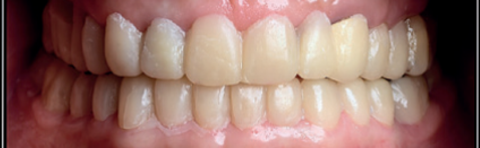
Badania obrazowe odgrywają coraz ważniejszą rolę w diagnostyce i monitorowaniu chorób stawów skroniowo‑żuchwowych. Celem pracy jest przedstawienie przebiegu rehabilitacji protetycznej w chorobie zwyrodnieniowej, przeprowadzonej w oparciu o diagnostykę radiologiczną na podstawie opisu przypadku 52‑letniego pacjenta z osteoartrozą lewego stawu skroniowo‑żuchwowego. Trwające 15 miesięcy leczenie wieloetapowe, zakończone pełną rekonstrukcją zgryzu, rozpoczęto w aktywnej fazie choroby, gdy głównym objawem u pacjenta był ból okolicy lewego stawu skroniowo‑żuchwowego. Przeprowadzono terapię przeciwbólową i przeciwzapalną lekami z grupy NLPZ oraz odwracalne leczenie zachowawcze w postaci szynoterapii. Po uzyskaniu stabilizacji, gdy choroba osiągnęła fazę nieaktywną, wykonano odbudowę protetyczną z wykorzystaniem licówek, nakładów, koron i mostów ceramicznych.
Hasła indeksowe: osteoartroza, staw skroniowo‑żuchwowy, tomografia wiązki stożkowej
Key words: osteoarthritis, temporomandibular joint, cone beam computed tomography (CBCT), magnetic resonance imaging (MRI)
Piśmiennictwo
1. Zarb G., Carlsson G.: Temporomandibular disorders: osteoarthritis. J. Orofac. Pain, 1999, 4, 13, 295-306.
2. Rasmussen O.C.: Clinical findings during the course of temporomandibular arthropathy. Scand. J. Dent. Res., 1981, 89, 3, 283-288.
3. Schmitter M., Kress B., Rammelsberg P.: Temporomandibular joint pathosis in patients with myofascial pain: a comparative analysis of magnetic resonance imaging and a clinical examination based on a specific set of criteria. Oral Surg. Oral Med. Oral Pathol. Oral Radiol. Endod., 2004, 97, 3, 318-324.
4. Manfredini D., Guarda-Nardini L.: Agreement between Research Diagnostic Criteria for Temporomandibular Disorders and magnetic resonance diagnoses of temporomandibular disc displacement in a patient population. Int. J. Oral Maxillofac. Surg., 2008, 37, 7, 612-616.
5. McLaren E., Culp L.: Smile analysis – the Photoshop smile design technique: Part I. J. Esthet. Dent., 2013, 29, 1, 94-108.
6. Culp L., McLaren E., Swann L.: Smile analysis–converting digital designs to the final smile: Part II. J. Esthet. Dent., 2013, 29, 2, 98-108.
7. Mejersjo C.: Therapeutic and prognostic considerations in TMJ osteoarthrosis: a literature review and a long-term study in 11 subjects. Cranio, 1987, 5, 1, 69-78.
8. Kalladka M. i wsp.: Temporomandibular joint osteoarthritis: diagnosis and long-term conservative management: a topic review. J. Indian Prosthodont. Soc., 2014, 14, 1, 6-15.
9. de Leeuw R. i wsp.: Symptoms of temporomandibular joint osteoarthrosis and internal derangement 30 years after non-surgical treatment. Cranio, 1995, 13, 2, 81-88.
10. deLeeuw R. i wsp.: Radiographic signs of temporomandibular joint osteoarthrosis and internal derangement 30 years after nonsurgical treatment. Oral Surg. Oral Med. Oral Pathol. Oral Radiol. Endod., 1995, 79, 3, 382-392.
11. Bansal M.: Addressing the Gaps: Therapeutic and Prognostic Considerations in Temporomandibular Joint Osteoarthrosis. J. Otolaryngol. ENT Res., 2017, 8,6, 00266. DOI: 10.15406/joentr.2017.08.00266.
12. Tamini D., Hatcher D.C.: Specialty Imaging: Temporomandibular Joint. Wyd. 1. Elsevier, 2016.
13. Schiffman E. i wsp.: Diagnostic Criteria for Temporomandibular Disorders (DC/TMD) for Clinical and Research Applications: recommendations of the International RDC/TMD Consortium Network and Orofacial Pain Special Interest Group. J. Oral Facial Pain Headache, 2014, 28, 1, 6-27.
14. Hunter A., Kalathingal S.: Diagnostic imaging for temporomandibular disorders and orofacial pain. Dent. Clin. North Am., 2013, 57, 3, 405-418.
15. Mawani F. i wsp.: Condylar shape analysis using panoramic radiography units and conventional tomography. Oral Surg. Oral Med. Oral Pathol. Oral Radiol. Endod., 2005, 99, 341-348.
16. Schmitter M. i wsp.: Assessment of the reliability and validity of panoramic imaging for assessment of mandibular condyle morphology using both MRI and clinical examination as the gold standard. Oral Surg. Oral Med. Oral Pathol. Oral Radiol. Endod., 2006, 102, 220-224.
17. Fallon S.D., Fritz G.W., Laskin D.M.: Panoramic imaging of the temporomandibular joint: an experimental study using cadaveric skulls. J. Oral Maxillofac. Surg., 2006, 64, 223-229.
18. Crow H.C. i wsp.: The utility of panoramic radiography in temporomandibular joint assessment. Dentomaxillofac. Radiol., 2005, 34, 2, 91-95.
19. Westesson P.L.: Structural hard-tissue changes in temporomandibular joints with internal derangement. Oral Surg. Oral Med. Oral Pathol., 1985, 59, 220-224.
20. Ahmad M. i wsp.: Research diagnostic criteria for temporomandibular disorders (RDC/TMD): development of image analysis criteria and examiner reliability for image analysis. Oral Surg. Oral Med. Oral Pathol. Oral Radiol. Endod., 2009, 107, 844-860.
21. Talmaceanu D. i wsp.: Imaging modalities for temporomandibular joint disorders: an update. Clujul Med., 2018, 91, 3, 280-287.
22. Honey O.B. i wsp.: Accuracy of cone-beam computed tomography imaging of the temporomandibular joint: comparisons with panoramic radiology and linear tomography. Am. J. Orthod. Dentofacial Orthop., 2007,132, 429-438.
23. Watanabe H. i wsp.: A comparative study for spatial resolution and subjective image characteristics of a multi-slice CT and a cone-beam CT for dental use. Eur. J. Radiol., 2011, 77, 397-402.
24. Zinman E.J., White S.C., Tetradis S.: Legal considerations in the use of cone beam computer tomography imaging. J. Calif. Dent. Assoc., 2010, 38, 1, 49-56.
25. Ludlow J.B., Ivanovic M.: Comparative dosimetry of dental CBCT devices and 64-slice CT for oral and maxillofacial radiology. Oral Surg. Oral Med. Oral Pathol. Oral Radiol. Endod., 2008, 106, 106-114.
26. Ludlow J.B., Davies-Ludlow L.E., White S.C.: Patient risk related to common dental radiographic examinations: the impact of 2007 International Commission on radiological protection recommendations regarding dose calculation. J. Am. Dent. Assoc., 2008, 139, 1237-1243.
27. Ludlow J.B.: A manufacturer’s role in reducing the dose of cone beam computed tomography examinations: effect of beam filtration. Dentomaxillofac. Radiol., 2011, 40, 115-122.
28. Scarfe W.C. i wsp.: Use of cone beam computed tomography in endodontics. Int. J. Dent., 2009, 1-20.
29. Barghan S., Tetradis S., Mallya S.: Application of cone beam computed tomography for assessment of the temporomandibular joints. Aust. Dent. J., 2012, 57, supl. 1, 109-118.
30. Szkutnik J., Litko M., Różyło-Kalinowska I.: Rola diagnostyki obrazowej w dysfunkcji narządu żucia. Mag. Stomatol., 2018, 5, 78-81.
31. Tomas X. i wsp.: MR imaging of temporomandibular joint dysfunction: a pictorial review. Radiographics, 2006, 26, 3, 765-781.
32. Sawada K. i wsp.: Diagnostic reliability of 3.0-T MRI for detecting osseous abnormalities of the temporomandibular joint. J. Oral Sci., 2018, 60, 1, 137-141.
33. Ma R-H. i wsp.: Application of fused image in detecting abnormalities of temporomandibular joint. Dentomaxillofac. Radiol., 2019, 48, 3, 20180129.
34. Al-Saleh M.A. i wsp.: MRI alone versus MRI-CBCT registered images to evaluate temporomandibular joint internal derangement. Oral Surg. Oral Med. Oral Pathol. Oral Radiol., 2016, 122, 638-645.
35. Habashi H. i wsp.: Dynamic high-resolution sonography compared to magnetic resonance imaging for diagnosis of temporomandibular joint disk displacement. J. Ultrasound Med., 2015, 34, 75-82.


