

Dostęp do tego artykułu jest płatny.
Zapraszamy do zakupu!
Cena: 12.50 PLN (z VAT)
Kup artykuł
Po dokonaniu zakupu artykuł w postaci pliku PDF prześlemy bezpośrednio pod twój adres e-mail.
Streszczenie
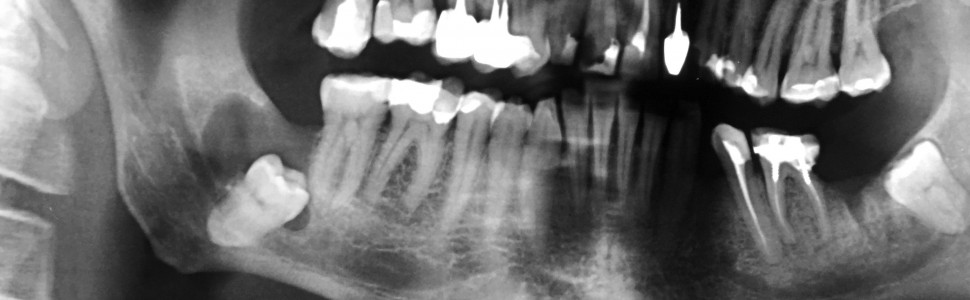
Torbiel zawiązkowa jest jedną z najczęstszych zmian w obszarze szczękowo‑twarzowym. Zazwyczaj jest związana z zatrzymanym zębem trzonowym trzecim żuchwy. Pacjenci najczęściej nie zgłaszają żadnych dolegliwości, gdyż rozwój zmiany jest przeważnie bezobjawowy. Torbiel zawiązkowa jest wykrywana przypadkowo na przeglądowych zdjęciach rentgenowskich. Całkowite wyłuszczenie torbieli jest postępowaniem z wyboru. W pracy przedstawiono przypadek 51 ‑letniego pacjenta z torbielą zawiązkową żuchwy związaną z zatrzymanym zębem 48.
Abstract
Dentigerous cyst is one of the most common lesions in the maxilla ‑facial area. It is usually associated with impacted mandibular third molars. Patients usually do not report any complaints, because the growth of the lesion is mostly asymptomatic. Dentigerous cyst is detected accidentally on routine radiographic images. Complete enucleation of the cyst is the treatment of choice. The study presents the case of 51 ‑year ‑old male patient with dentigerous cyst of mandible related with impacted tooth 48.
Hasła indeksowe: torbiel zawiązkowa, wyłuszczenie, żuchwa
Key words: dentigerous cyst, enucleation, mandible
PIŚMIENNICTWO
1. Kaczmarzyk T. i wsp.: Torbiele obszaru szczękowo ‑twarzowego. Wyd. Kwintesencja. Warszawa 2015, 58‑68.
2. Stelmach R., Osica P., Janas A.: Large dentigerous cysts of the mandible in a 46 ‑year ‑old
patient – a case report. J. Educ. Health Sport, 2017, 7, 1, 133‑140. DOI http://dx.doi.org/10.5281/zenodo.234255, http://ojs.ukw.edu.pl/index.php/johs/article/view/4157
3. Błochowiak K. i wsp.: Rare case of a huge odontogenic cyst of the mandible. Dent. Forum, 2013, 41, 144‑149.
4. Sridevi K. i wsp.: Dentigerous cysts of maxillofacial region – clinical, radiographic and biochemical analysis. Kathmandu Univ. Med. J., 2015, 13, 49, 8‑11.
5. Lin H.P. i wsp.: A clinicopathological study of 338 dentigerous cysts. J. Oral Pathol. Med., 2013, 42, 6, 462‑467.
6. Avelar R.L. i wsp.: Odontogenic cysts: a clinicopathological study of 507 cases. J. Oral Sci., 2009, 51, 4, 581‑586.
7. Deana N.F., Alves N.: Cone beam CT in diagnosis and surgical planning of dentigerous cyst. Case Rep. Dent., 2017, 2017, 7956041. Epub 2017, Feb 15.
8. Asnani S. i wsp.: Dentigerous cyst with an impacted third molar obliterating complete maxillary sinus. Indian J. Dent. Res., 2012, 23, 833‑835.
9. Gendviliene I. i wsp.: Conservative management of large mandibular dentigerous cysts with a novel approach for follow up: two case reports. Stomatologija, 2017, 19, 1, 24‑32.
10. Zapała ‑Pośpiech A. i wsp.: Malignant transformation in the course of a dentigerous cyst: a problem for a clinician and a pathologist. Considerations based on a case report. Pol. J. Pathol., 2013, 64, 1, 64‑68.
11. Pinto A.S. i wsp.: Value of magnetic resonance imaging for diagnosis of dentigerous cyst. Case Rep. Dent., 2016, 2016, 2806235. Epub. 2016 Sep 27.
Gawęda


