

Dostęp do tego artykułu jest płatny.
Zapraszamy do zakupu!
Cena: 12.50 PLN (z VAT)
Kup artykuł
Po dokonaniu zakupu artykuł w postaci pliku PDF prześlemy bezpośrednio pod twój adres e-mail.
The use of ultrasonography in monitoring the progress of treatment of functional disorders of the masticatory system
Marcin Wójcicki, Jacek Szkutnik, Ingrid Różyło-Kalinowska
Streszczenie
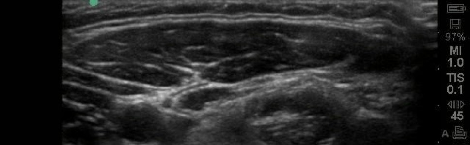
Zaburzenia czynnościowe narządu żucia (ZCNŻ) stanowią niejednorodną grupę schorzeń, w skład której wchodzą zarówno patologie dotyczące stawów skroniowo-żuchwowych (ssż), jak i mięśni narządu żucia. Wybór najlepszej metody leczenia jest niełatwym zadaniem dla lekarza dentysty i często dokonuje się go, analizując skuteczność prowadzonego leczenia. Niezmiernie istotna jest wówczas możliwość obiektywnego ocenienia postępów terapii. Do tego celu można zastosować badanie ultrasonograficzne (USG), które pozwala na ocenę struktur ssż oraz mięśni narządu żucia. Do zalet USG zaliczamy dużą dostępność, brak działań niepożądanych, wysoką rozdzielczość obrazu, możliwość obrazowania w czasie rzeczywistym, brak specjalnych procedur przygotowawczych oraz stosunkowo niski koszt badania.
Celem pracy było przedstawienie możliwości diagnozowania i monitorowania postępów leczenia za pomocą ultrasonografii u pacjentów z zaburzeniami czynnościowymi narządu żucia. W przypadku badania mięśni z wykorzystaniem USG jesteśmy w stanie ocenić ich grubość oraz budowę wewnętrzną, które korelują z mięśniowymi dolegliwościami bólowymi. W zaburzeniach wewnątrzstawowych na obrazie USG jesteśmy w stanie ocenić szczelinę stawu, pozycję krążka, występowanie adhezji, szerokość torebki stawowej, obrzęk stawu oraz erozję wyrostka kłykciowego. Zastosowanie USG w diagnostyce i monitorowaniu leczenia ZCNŻ wymaga przeprowadzenia dalszych rozbudowanych badań i standaryzacji tej metody obrazowania. Biorąc pod uwagę niski koszt, nieinwazyjność, brak działań niepożądanych, brak przeciwwskazań oraz krótki czas badania – w porównaniu z MR i CBCT – sugeruje się coraz częstsze sięganie po USG w praktyce stomatologicznej.
Abstract
Temporomandibular disorders (TMD) are a heterogenous group of diseases which concern both temporomandibular joints (TMJ) and masticatory muscles. Choosing the best method of treatment is not an easy task for a dentist and it is often done by assessing the effectiveness of the therapy. The ability to objectively assess the progress of treatment is extremely important. Ultrasonography (US) can be used for this purpose, which allows the assessment of the TMJ and masticatory organs structures. The advantages of US include high availability, no adverse effects, high image resolution, real-time imaging, no special preparatory procedures and a relatively low cost of testing. The aim of the study was to present the possibility of diagnosing and monitoring the progress of the treatment using ultrasonography in patients with functional disorders of the masticatory system. In the case of muscle examination using ultrasound it is possible to assess their thickness and internal structure, which correlate with muscular pain. In intra-articular disorders in the ultrasound image, it is possible to assess the joint fissure, the position of the disc, the occurrence of adhesion, the width of the joint capsule, swelling of the joint and erosion of the condylar process. The use of US in the diagnosis and monitoring of TMD treatment requires further extensive research and standardization of this imaging method. Given the low cost, non-invasiveness, no side effects, no contraindications and short duration of the examination it is suggested to use US in dental practice more often.
Hasła indeksowe: ultrasonografia, zaburzenia czynnościowe narządu żucia
Key words: ultrasonography, temporomandibular disorders
PIŚMIENNICTWO
1. Okeson J.P.: Leczenie dysfunkcji skroniowo-żuchwowych i zaburzeń zwarcia. Wydawnictwo Czelej, Lublin 2018, 147-186.
2. Bakalczuk M. i wsp.: Intra-rater reliability of TMJ joint vibration – a pilot study. EJMT, 2017, 1, 8-12.
3. Bakalczuk M. i wsp.: Analysis of TMJ joint vibration with and without silicon splints in patients with temporomandibular joint hypermobility – a clinical study. EJMT, 2018, 2, 12-16.
4. Różyło-Kalinowska I., Różyło T.K.: Współczesna radiologia stomatologiczna. Wydawnictwo Czelej, Lublin 2012, 52-57.
5. Serra M.D., Duarte Gavião M.B., dos Santos Uchôa M.N.: The use of ultrasound in the investigation of the muscles of mastication. Ultrasound Med. Biol., 2008, 34, 1875-1884.
6. Vanneuville G. i wsp.: The anatomical basis for ultrasonography of the lateral pterygoid muscle and the infratemporal fossa. Surg. Radiol. Anat., 1994, 16, 57-61.
7. Ariji Y. i wsp.: Masseter muscle sonographic features as indices for evaluating efficacy of massage treatment. Oral Surg. Oral Med. Oral Pathol. Oral Radiol. Endod., 2010, 110, 517-526.
8. Kiliaridis S., Engvall M., Tzakis M.G.: Ultrasound imaging of the masseter muscle in myotonic dystrophy patients. J. Oral Rehabil., 1995, 22, 619-625.
9. Ariji Y. i wsp.: Ultrasonographic features of the masseter muscle in female patients with temporomandibular disorder associated with myofascial pain. Oral Surg. Oral Med. Oral Pathol. Oral Radiol. Endod., 2004, 98, 337-341.
10. Emshoff R. i wsp.: Reliability and temporal variation of masseter muscle thickness measurements utilizing ultrasonography. J. Oral Rehabil., 2003, 30, 1168-1172.
11. Emshoff R., Bertram S., Strobl H.: Ultrasonographic cross-sectional characteristics of muscles of the head and neck. Oral Surg. Oral Med. Oral Pathol. Oral Radiol. Endod., 1999, 87, 93-106.
12. Goller Bulut D., Avci F., Özcan G.: Ultrasonographic evaluation of jaw elevator muscles in young adults with bruxism and with and without attrition-type tooth wear: A pilot study. Cranio., 2018, August, 1-8.
13. Bertram S. i wsp.: Effect of scanning level and muscle condition on ultrasonographic cross-sectional measurements of the anterior masseter muscle. J. Oral Rehabil., 2003, 30, 430-435.
14. Busato A. i wsp.: Strain analysis of masseter muscle by ultrasound. J. Biol. Regul. Homeost. Agents, 2015, 29, 74-81.
15. Baba I.A. i wsp.: TMJ imaging: a review. Int. J. Contemp. Med. Res., 2016, 3, 2253-2256.
16. Sakhavalkar P., Bhoosreddy A., Kotwal H.: Assessment and comparison of the capsular width of temporomandibular joint on ultrasonography and magnetic resonance imaging. J. Indian Acad. Oral Med. Radiol., 2016, 28, 351.
17. Różyło-Kalinowska I., Orhan K.: Imaging of the Temporomandibular Joint. Springer Berlin Heidelberg, New York 2018, 113-152.
18. Siva Kalyan U., Moturi K., Padma Rayalu K.: The role of ultrasound in diagnosis of temporomandibular joint disc displacement: a case–control study. J. Maxillofac. Oral Surg., 2018, 17, 383-388.
19. Azlağ Pekince K., Çağlayan F., Pekince A.: The efficacy and limitations of USI for diagnosing TMJ internal derangements. Oral Radiol., 2019, 4, doi:10.1007/s11282-019-00376-3
20. Chalkoo A., Ahmad M., Naikoo F.: Magnetic resonance imaging and ultrasonography in the diagnosis of temporomandibular joint internal derangements: A comparative study. J. Indian Acad. Oral Med. Radiol., 2015, 27, 198.
21. Lanni S. i wsp.: Towards a role of ultrasound in children with juvenile idiopathic arthritis. Rheumatology, 2013, 52, 413-420.
22. Wakefield R.J. i wsp.: The value of sonography in the detection of bone erosions in patients with rheumatoid arthritis: A comparison with conventional radiography. Arthritis Rheum., 2000, 43, 2762-2770.
23. Telkar S. i wsp.: Evaluation of occlusal splint therapy in temporomandibular joint disorder patients using real-time ultrasonography. J. Investig. Clin. Dent., 2010, 1, 96-100.
24. Bertram S. i wsp.: Effect of stabilization-type splints on the asymmetry of masseter muscle sites during maximal clenching. J. Oral Rehabil., 2002, 29, 447-451.
25. Park M.Y., Ahn K.Y., Jung D.S.: Botulinum toxin type A treatment for contouring of the lower face. Dermatol. Surg., 2003, 29, 477-483.
26. Ahn J., Horn C., Blitzer A.: Botulinum toxin for masseter reduction in Asian patients. Arch. Facial Plast. Surg., 2004, 6, 188-191.
27. Kubo K. i wsp.: Outer shape changes of human masseter with contraction by ultrasound morphometry. Arch. Oral Biol., 2006, 51, 146-153.
Marcin Wójcicki, Jacek Szkutnik, Ingrid Różyło-Kalinowska
Streszczenie
Zaburzenia czynnościowe narządu żucia (ZCNŻ) stanowią niejednorodną grupę schorzeń, w skład której wchodzą zarówno patologie dotyczące stawów skroniowo-żuchwowych (ssż), jak i mięśni narządu żucia. Wybór najlepszej metody leczenia jest niełatwym zadaniem dla lekarza dentysty i często dokonuje się go, analizując skuteczność prowadzonego leczenia. Niezmiernie istotna jest wówczas możliwość obiektywnego ocenienia postępów terapii. Do tego celu można zastosować badanie ultrasonograficzne (USG), które pozwala na ocenę struktur ssż oraz mięśni narządu żucia. Do zalet USG zaliczamy dużą dostępność, brak działań niepożądanych, wysoką rozdzielczość obrazu, możliwość obrazowania w czasie rzeczywistym, brak specjalnych procedur przygotowawczych oraz stosunkowo niski koszt badania.
Celem pracy było przedstawienie możliwości diagnozowania i monitorowania postępów leczenia za pomocą ultrasonografii u pacjentów z zaburzeniami czynnościowymi narządu żucia. W przypadku badania mięśni z wykorzystaniem USG jesteśmy w stanie ocenić ich grubość oraz budowę wewnętrzną, które korelują z mięśniowymi dolegliwościami bólowymi. W zaburzeniach wewnątrzstawowych na obrazie USG jesteśmy w stanie ocenić szczelinę stawu, pozycję krążka, występowanie adhezji, szerokość torebki stawowej, obrzęk stawu oraz erozję wyrostka kłykciowego. Zastosowanie USG w diagnostyce i monitorowaniu leczenia ZCNŻ wymaga przeprowadzenia dalszych rozbudowanych badań i standaryzacji tej metody obrazowania. Biorąc pod uwagę niski koszt, nieinwazyjność, brak działań niepożądanych, brak przeciwwskazań oraz krótki czas badania – w porównaniu z MR i CBCT – sugeruje się coraz częstsze sięganie po USG w praktyce stomatologicznej.
Abstract
Temporomandibular disorders (TMD) are a heterogenous group of diseases which concern both temporomandibular joints (TMJ) and masticatory muscles. Choosing the best method of treatment is not an easy task for a dentist and it is often done by assessing the effectiveness of the therapy. The ability to objectively assess the progress of treatment is extremely important. Ultrasonography (US) can be used for this purpose, which allows the assessment of the TMJ and masticatory organs structures. The advantages of US include high availability, no adverse effects, high image resolution, real-time imaging, no special preparatory procedures and a relatively low cost of testing. The aim of the study was to present the possibility of diagnosing and monitoring the progress of the treatment using ultrasonography in patients with functional disorders of the masticatory system. In the case of muscle examination using ultrasound it is possible to assess their thickness and internal structure, which correlate with muscular pain. In intra-articular disorders in the ultrasound image, it is possible to assess the joint fissure, the position of the disc, the occurrence of adhesion, the width of the joint capsule, swelling of the joint and erosion of the condylar process. The use of US in the diagnosis and monitoring of TMD treatment requires further extensive research and standardization of this imaging method. Given the low cost, non-invasiveness, no side effects, no contraindications and short duration of the examination it is suggested to use US in dental practice more often.
Hasła indeksowe: ultrasonografia, zaburzenia czynnościowe narządu żucia
Key words: ultrasonography, temporomandibular disorders
PIŚMIENNICTWO
1. Okeson J.P.: Leczenie dysfunkcji skroniowo-żuchwowych i zaburzeń zwarcia. Wydawnictwo Czelej, Lublin 2018, 147-186.
2. Bakalczuk M. i wsp.: Intra-rater reliability of TMJ joint vibration – a pilot study. EJMT, 2017, 1, 8-12.
3. Bakalczuk M. i wsp.: Analysis of TMJ joint vibration with and without silicon splints in patients with temporomandibular joint hypermobility – a clinical study. EJMT, 2018, 2, 12-16.
4. Różyło-Kalinowska I., Różyło T.K.: Współczesna radiologia stomatologiczna. Wydawnictwo Czelej, Lublin 2012, 52-57.
5. Serra M.D., Duarte Gavião M.B., dos Santos Uchôa M.N.: The use of ultrasound in the investigation of the muscles of mastication. Ultrasound Med. Biol., 2008, 34, 1875-1884.
6. Vanneuville G. i wsp.: The anatomical basis for ultrasonography of the lateral pterygoid muscle and the infratemporal fossa. Surg. Radiol. Anat., 1994, 16, 57-61.
7. Ariji Y. i wsp.: Masseter muscle sonographic features as indices for evaluating efficacy of massage treatment. Oral Surg. Oral Med. Oral Pathol. Oral Radiol. Endod., 2010, 110, 517-526.
8. Kiliaridis S., Engvall M., Tzakis M.G.: Ultrasound imaging of the masseter muscle in myotonic dystrophy patients. J. Oral Rehabil., 1995, 22, 619-625.
9. Ariji Y. i wsp.: Ultrasonographic features of the masseter muscle in female patients with temporomandibular disorder associated with myofascial pain. Oral Surg. Oral Med. Oral Pathol. Oral Radiol. Endod., 2004, 98, 337-341.
10. Emshoff R. i wsp.: Reliability and temporal variation of masseter muscle thickness measurements utilizing ultrasonography. J. Oral Rehabil., 2003, 30, 1168-1172.
11. Emshoff R., Bertram S., Strobl H.: Ultrasonographic cross-sectional characteristics of muscles of the head and neck. Oral Surg. Oral Med. Oral Pathol. Oral Radiol. Endod., 1999, 87, 93-106.
12. Goller Bulut D., Avci F., Özcan G.: Ultrasonographic evaluation of jaw elevator muscles in young adults with bruxism and with and without attrition-type tooth wear: A pilot study. Cranio., 2018, August, 1-8.
13. Bertram S. i wsp.: Effect of scanning level and muscle condition on ultrasonographic cross-sectional measurements of the anterior masseter muscle. J. Oral Rehabil., 2003, 30, 430-435.
14. Busato A. i wsp.: Strain analysis of masseter muscle by ultrasound. J. Biol. Regul. Homeost. Agents, 2015, 29, 74-81.
15. Baba I.A. i wsp.: TMJ imaging: a review. Int. J. Contemp. Med. Res., 2016, 3, 2253-2256.
16. Sakhavalkar P., Bhoosreddy A., Kotwal H.: Assessment and comparison of the capsular width of temporomandibular joint on ultrasonography and magnetic resonance imaging. J. Indian Acad. Oral Med. Radiol., 2016, 28, 351.
17. Różyło-Kalinowska I., Orhan K.: Imaging of the Temporomandibular Joint. Springer Berlin Heidelberg, New York 2018, 113-152.
18. Siva Kalyan U., Moturi K., Padma Rayalu K.: The role of ultrasound in diagnosis of temporomandibular joint disc displacement: a case–control study. J. Maxillofac. Oral Surg., 2018, 17, 383-388.
19. Azlağ Pekince K., Çağlayan F., Pekince A.: The efficacy and limitations of USI for diagnosing TMJ internal derangements. Oral Radiol., 2019, 4, doi:10.1007/s11282-019-00376-3
20. Chalkoo A., Ahmad M., Naikoo F.: Magnetic resonance imaging and ultrasonography in the diagnosis of temporomandibular joint internal derangements: A comparative study. J. Indian Acad. Oral Med. Radiol., 2015, 27, 198.
21. Lanni S. i wsp.: Towards a role of ultrasound in children with juvenile idiopathic arthritis. Rheumatology, 2013, 52, 413-420.
22. Wakefield R.J. i wsp.: The value of sonography in the detection of bone erosions in patients with rheumatoid arthritis: A comparison with conventional radiography. Arthritis Rheum., 2000, 43, 2762-2770.
23. Telkar S. i wsp.: Evaluation of occlusal splint therapy in temporomandibular joint disorder patients using real-time ultrasonography. J. Investig. Clin. Dent., 2010, 1, 96-100.
24. Bertram S. i wsp.: Effect of stabilization-type splints on the asymmetry of masseter muscle sites during maximal clenching. J. Oral Rehabil., 2002, 29, 447-451.
25. Park M.Y., Ahn K.Y., Jung D.S.: Botulinum toxin type A treatment for contouring of the lower face. Dermatol. Surg., 2003, 29, 477-483.
26. Ahn J., Horn C., Blitzer A.: Botulinum toxin for masseter reduction in Asian patients. Arch. Facial Plast. Surg., 2004, 6, 188-191.
27. Kubo K. i wsp.: Outer shape changes of human masseter with contraction by ultrasound morphometry. Arch. Oral Biol., 2006, 51, 146-153.


