

Dostęp do tego artykułu jest płatny.
Zapraszamy do zakupu!
Po dokonaniu zakupu artykuł w postaci pliku PDF prześlemy bezpośrednio pod twój adres e-mail.
ARTYKUŁ ORYGINALNY
Analiza długości szwu podniebiennego przy użyciu tomografii komputerowej wiązki stożkowej
Analysis of the length of the palatal suture using cone beam computed tomography
Olga Zajączkowska, Klaudia Lulek, Magdalena Piskórz, Ingrid Różyło-Kalinowska
Streszczenie
Wstęp: Szew podniebienny łączy wyrostki podniebienne szczęk, które razem tworzą podniebienie twarde. Najczęściej jest on oceniany podczas planowania leczenia ortodontycznego oraz w zakresie chirurgii ortognatycznej. Ocena szwu może być wykorzystywana podczas leczenia nasilonych zwężeń szczęk, przodozgryzów rzekomych, morfologicznego niedorozwoju szczęk czy rozszczepów. Diagnostyka obrazowa z wykorzystaniem obrazowania wolumetrycznego (CBCT) jest kluczowym czynnikiem przyczyniającym się do prawidłowego rozpoznania ortodontycznego w skomplikowanych przypadkach. Dzięki zastosowaniu badania CBCT możemy obrazować przebieg, długość i kształt szwu podniebiennego oraz planować ewentualne leczenie w sytuacji, gdy stwierdzane są zaburzenia.
Cel pracy: Celem pracy była analiza długości szwu podniebiennego pośrodkowego po wyznaczeniu punktów referencyjnych u pacjentów w wieku 7-20 lat z uwzględnieniem płci przy użyciu CBCT.
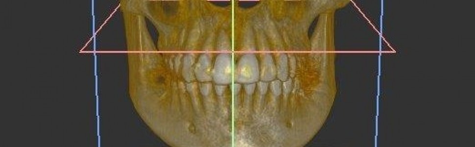
Materiały imetody: Materiał stanowiło 90 badań CBCT wyeksportowanych z bazy danych pacjentów Zakładu Rentgenodiagnostyki Stomatologicznej i Szczękowo-Twarzowej Uniwersytetu Medycznego w Lublinie, z aparatu VistaVox S firmy Dürr Dental (Niemcy). W programie Vista Soft (Dürr Dental) dokonano pomiaru długości szwu podniebiennego, następnie wyniki poddano analizie statystycznej opisowej.
Wyniki: Nie stwierdzono znacznej zależności i różnicy między płcią a długością szwu podniebiennego. Rozpatrując poszczególne wyniki długości szwu podniebiennego, stwierdzono jego dodatnią zależność z wiekiem, natomiast występujące w danych grupach wiekowych znaczne odchylenia od pozostałych wartości zmodyfikowały ostateczny bilans. U płci męskiej zauważono tendencję wzrostową, a dodatkowo w starszej grupie pacjentów występowały wyższe wartości mierzonej struktury anatomicznej.
Abstract
Introduction: The palatal suture connects the palatal processes of the maxilla, forming the hard palate. It is commonly assessed during orthodontic treatment planning and in the context of orthognathic surgery. The evaluation of the suture can be utilized in the treatment of severe maxillary constrictions, pseudo-class III malocclusions, morphological underdevelopment of the maxilla or clefts. Medical imaging using volumetric imaging, such as cone-beam computed tomography (CBCT), is acrucial factor contributing to accurate orthodontic diagnosis in complex cases. Through CBCT examinations, the course, length, and shape of the palatal suture can be visualized, aiding in treatment planning when abnormalities are identified.
Objective: The aim of this study was to analyze the length of the midline palatal suture after determining reference points in patients aged 7-20 years, taking gender into account, using CBCT.
Materials and methods: The material consisted of 90 CBCT scans exported from the database of the Department of Dental Radiology and Maxillofacial Radiology at the Medical University of Lublin, obtained with the VistaVox S device from Dürr Dental (Germany). The Vista Soft program (Dürr Dental) was used to measure the length of the palatal suture, and the results were subjected to descriptive statistical analysis.
Results: No signifi cant correlation or difference was found between gender and the length of the palatal suture. Examining individual results of the palatal suture length revealed apositive correlation with age. However, substantial deviations from other values within specific age groups modified the final balance. In the male group, agrowth trend was observed, with higher values of the measured anatomical structure in the older patient group.
Hasła indeksowe: szew podniebienny, długość szwu podniebiennego, CBCT
Key words: palatal suture, length of the palatal suture, CBCT
Piśmiennictwo
1. Shibusawa N, Endo Y, Morimoto N i wsp. Mathematical modeling of palatal suture pattern formation: morphological differences between sagittal and palatal sutures. Sci Rep. 2021; 11(1): 8995.
2. Proffit WR, Fields HW, Larson B i wsp. Contemporary Orthodontics, wyd. 6. Wrocław: Elsevier Ltd. Oxford; 2009, s. 242, 278.
3. Karłowska I. Zarys współczesnej ortodoncji. Podręcznik dla studentów i lekarzy dentystów, wyd. 4. Warszawa: PZWL; 2016, s. 165, 175, 240-241.
4. Zawiślak E. Wczesne efekty leczenia metodą dystrakcji przezpodniebiennej zwężeń szczęki u pacjentów dorosłych, [rozprawa doktorska]. Wrocław: Uniwersytet Medyczny im. Piastów Śląskich; 2019, s. 21-106.
5. Abrol V, Abrol K. CBCT: Third eye in dentistry. IJODR 2018; 4(1): 6-8.
6. Abizadeh N, Moles DR, O’Neill J i wsp. Digital versus plaster study models: how accurate and reproducible are they? J Orthod. 2012; 39(3): 151-159.
7. Arai Y, Tammisalo E, Iwai K i wsp. Development of a compact computed tomographic apparatus for dental use. Dentomaxillofac Radiol. 1999; 28(4): 245-248.
8. Garib DG, Yatabe MS, Ozawa TO i wsp. Alveolar bone morphology in patients with bilateral complete cleft lip and palate in the mixed dentition: cone beam computed tomography evaluation. Cleft Palate Cra niofac J. 2012; 49(2): 208-214.
9. Pauwels R. Cone beam CT for dental and maxillofacial imaging: dose matters. Radiat Prot Dosimetry. 2015; 165(1-4): 156-161.
10. De Grauwe A, Ayaz I, Shujaat S i wsp. CBCT in orthodontics: a systematic review on justification of CBCT in a paediatric population prior to orthodontic treatment. Eur J Orthod. 2019; 41(4): 381-389.
11. Hamada Y, Kondoh T, Noguchi K i wsp. Application of limited cone beam computed tomography to clinical assessment of alveolar bone grafting: a preliminary report. Cleft Palate Craniofac J. 2005; 42(2): 128-137.
12. Wörtche R, Hassfeld S, Lux CJ i wsp. Clinical application of cone beam digital volume tomography in children with cleft lip and palate. Dento maxillofacial Radiol. 2006; 35(2): 88-94.
13. Chae JM, Rogowski L, Mandair S i wsp. A CBCT Evaluation of Midpalatal Bone Density in Various Skeletal Patterns. Sensors (Basel). 2021; 21(23): 7812.
14. Shin H, Hwang CJ, Lee KJ i wsp. Predictors of midpalatal suture expansion by miniscrew-assisted rapid palatal expansion in young adults: A preliminary study. Korean J Orthod. 2019; 49(6): 360-371.
15. Ok G, Yilmaz BS, Aksoy DO i wsp. Maturity evaluation of orthodonti cally important anatomic structures with computed tomography. Eur J Orthod. 2021; 43(1): 8-14.
16. Savoldi F, Wong KK, Yeung AWK i wsp. Midpalatal suture maturation staging using cone beam computed tomography in patients aged be tween 9 to 21 years. Sci Rep. 2022; 12(1): 4318.
17. Haghanifar S, Mahmoudi S, Foroughi R i wsp. Assessment of midpalatal suture ossification using cone-beam computed tomography. Electron Physician. 2017; 9(3): 4035-4041.
18. Christovam IO, Lisboa CO, Vilani GNL i wsp. Tomographic analysis of midpalatal suture prior to rapid maxillary expansion. Dental Press J Or thod. 2021; 26(3): e2119300.
19. Manabe A, Ishida T, Kanda E i wsp. Evaluation of maxillary and mandibular growth patterns with cephalometric analysis based on cervical vertebral maturation: A Japanese cross-sectional study. PLoS One. 2022; 17(4): e0265272.
20. Papageorgiou SN, Kutschera E, Memmert S i wsp. Effectiveness of early orthopaedic treatment with headgear: a systematic review and meta--analysis. Eur J Orthod. 2017; 39(2): 176-187.
21. Çoban G, Öztürk T, Erdem ME i wsp. Effects of maxillary expansion and protraction on pharyngeal airway dimensions in relation to changes in head posture and hyoid position: A retrospective cohort study. J Orofac Orthop. 2022; 84(Suppl 3): 172-185.
22. Trevizan M, Filho PN, Franzolin SOB i wsp. Premaxilla: up to which age it remains separated from the maxilla by a suture, how often it occurs in children and adults, and possible clinical and therapeutic implications: Study of 1,138 human skulls. Dental Press J Orthod. 2018; 23(6): 16-29.
23. Colak O, Paredes NA, Elkenawy I i wsp. Tomographic assessment of palatal suture opening pattern and pterygopalatine suture disarticulation in the axial plane after midfacial skeletal expansion. Prog Orthod. 2020; 21(1): 21.


