

Dostęp do tego artykułu jest płatny.
Zapraszamy do zakupu!
Po dokonaniu zakupu artykuł w postaci pliku PDF prześlemy bezpośrednio pod twój adres e-mail.
STUDIA PRZYPADKÓW
Little molars, czyli strzonowaciałe przedtrzonowce
Little molars
Mariya Kubatska
Streszczenie
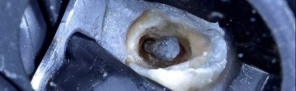
Każdy doświadczony endodonta marzy o przypadku leczenia trzykorzeniowego zęba przedtrzonowego w swoim endodontycznym portfolio. Co powoduje, że owa wariacja anatomiczna jest tak pożądana? Przede wszystkim to, że występuje bardzo rzadko. Po przeglądzie piśmiennictwa stwierdzono, że częstość występowania trzykorzeniowych zębów przedtrzonowych, nazywanych także little molars, wynosi 1-4% w przypadku pierwszych zębów przedtrzonowych oraz poniżej 1% w przypadku drugich zębów przedtrzonowych. W Polsce odsetek występowania trzykorzeniowych zębów przedtrzonowych w ogóle wynosi aż 9,2%. Leczenie trzykorzeniowych zębów przedtrzonowych należy do jednych z najbardziej wymagających i trudnych przypadków endodontycznych, nie tylko ze względu na bardzo rzadkie występowanie, ale i z uwagi na: trudności w diagnostyce, dostęp, instrumentację, odbudowę protetyczną, bardzo delikatną budowę korzeni. W pracy opisano trzy przypadki leczenia trzykorzeniowych zębów przedtrzonowych, w tym jeden opatrzono filmem.
Abstract
Every experienced endodontist dreams of having a three-rooted premolar treatment case in his endodontic portfolio. What makes this anatomical variation so desirable? First of all, it is very rare. After reviewing the literature, it was found that the incidence of three-rooted premolars, also called little molars, is 1-4% in the case of fi rst premolars and less than 1% in the case of second premolars. In Poland, the percentage of three-rooted premolars is as high as 9.2%. Treatment of three-rooted premolars is one of the most demanding and difficult endodontic cases, not only due to their very rare occurrence, but also due to: difficulties in diagnosis, access, instrumentation, prosthetic reconstruction, and very delicate structure of the roots. The paper describes three cases of treatment of three-rooted premolars, including one with a video.
Hasła indeksowe: zęby przedtrzonowe, little molars, trzykorzeniowy ząb przedtrzonowy, leczenie endodontyczne
Key words: premolars, little molars, three-rooted premolar, endodontic treatment
Film. Nagranie z badania CBCT.
Piśmiennictwo
- Carns EJ, Skidmore AE. Configurations and deviations of root canals of maxillary first premolars. Oral Surg Oral Med Oral Pathol. 1973; 36(6): 880-886.
- Vertucci FJ. Root canal anatomy of the human permanent teeth. Oral Surg Oral Med Oral Pathol. 1984; 58(5): 589-599.
- De Deus QD. Frequency, location and direction of the lateral, secondary and accessory canal. J Endod. 1975; 1: 361–366.
- Pecora JD, Saquy PC, Sousa Neto MD i wsp. Root form and canal anatomy of maxillary first premolars. Braz Dent J. 1992; 2: 87-94.
- Neelakantan P, Subbarao C, Ahuja R i wsp. Root and canal morphology of Indian maxillary premolars by a modified root canal staining technique. Odontology. 2011; 99: 18-21.
- Gupta S, Sinha DJ, Gowhar O i wsp. Root and canal morphology of maxillary first premolar teeth in North Indian population using clearing technique: An in vitro study. J Conserv Dent. 2015; 18: 232–236.
- Kartal N, Ozcelik B, Cimilli H. Root canal morphology of maxillary premolars. J Endod. 1998; 24: 417-419.
- Ozcan E, Colak H, Hamidi MM. Root and canal morphology of maxillary first premolars in a Turkish population. JDS. 2012; 7(4): 390-394.
- Lipski M. Root surface temperature rises in vitro during root canal obturation using hybrid and microseal techniques. J Endod. 2005; 31(4): 297-300.
- Walker RT. Root form and canal anatomy of maxillary first premolars in a southern Chinese population. Endod Dent Traumatol. 1987; 3: 130-134.
- Cheng XL, Weng YL. Observation of the roots and root canals of 442 maxillary first premolars. Shanghai Kou Qiang Yi Xue. 2008;17(5): 525-528.
- Tian YY, Guo B, Zhang R i wsp. Root and canal morphology of maxillary first premolars in a Chinese subpopulation evaluated using cone-beam computed tomography. Inter Endod J. 2012; 45: 996–1003.
- Loh HS. Root morphology of the maxillary first premolar in Singaporeans. Aust Dent J. 1998; 43: 399-402.
- Ahmad IA, Alenezi MA. Root and root canal morphology of maxillary first premolars. A literature review and clinical considerations. J Endod. 2016; 42(6): 861-872.
- Wolf TG, Kozaczek C, Siegrist M i wsp. An ex vivo study of root canal system configuration and morphology of 115 maxillary first premolars. J Endod. 2020; 46(6): 794-800.
- Hartmann RC, Baldasso FE, Stürmer CP i wsp. Clinically relevant dimensions of 3-rooted maxillary premolars obtained via high-resolution computed tomography. J Endod. 2013; 39(12): 1639-1645.


