

Dostęp do tego artykułu jest płatny.
Zapraszamy do zakupu!
Cena: 12.50 PLN (z VAT)
Kup artykuł
Po dokonaniu zakupu artykuł w postaci pliku PDF prześlemy bezpośrednio pod twój adres e-mail.
MS 2019; 10: 42-46.
Streszczenie
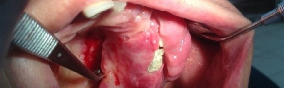
Zlokalizowane zmiany rozrostowe dziąseł należą do najczęstszych zmian proliferacyjnych stwierdzanych w jamie ustnej. Stanowią one obszerną grupę, obejmującą zarówno guzy łagodne, jak i nowotwory złośliwe. Przykładem zmiany łagodnej, pojawiającej się w obrębie dziąseł, jest włókniak kostniwno-kostniejący (cemento-ossifying fibroma – COF). Do 2017 roku był on klasyfikowany jako włókniak kostniejący obwodowy (peripheral ossifying fibroma – POF). Stanowi 9,6% wszystkich przypadków rozrostów dziąsłowych, występuje zwykle u osób w 2. i 3. dekadzie życia, z przewagą kobiet. Najczęściej lokalizuje się w przednim odcinku szczęki lub żuchwy. Ma charakter reaktywny, nienowotworowy, wywodzi się z więzadeł ozębnej. Występowanie tej zmiany wiąże się z obecnością czynników drażniących, takich jak kamień nazębny, wadliwe uzupełnienia protetyczne czy wypełnienia zachowawcze. Ostateczna diagnoza powinna zostać potwierdzona badaniem histopatologicznym, które ujawnia obecność ognisk kostnienia w tkance łącznej. Chirurgiczne wycięcie guza jest leczeniem z wyboru. Wznowa może występować w około 20% przypadków.
W artykule opisano przypadek 48-letniej kobiety, która zgłosiła się do Zakładu Chirurgii Stomatologicznej Warszawskiego Uniwersytetu Medycznego w celu konsultacji i leczenia dużej uszypułowanej zmiany rozrostowej o charakterze COF w okolicy zęba 26. W kolejnych częściach artykułu przedstawiono kliniczny, radiologiczny i histologiczny opis tego guza.
Abstract
Local gingival enlargements are the most frequent type of proliferative lesions in the oral cavity. They make up a wide group, which includes both benign and malignant tumors. One of benign lesions affecting gingival area is cemento-ossifying fibroma – COF.
Cemento-ossifying fibroma was classified as peripheral ossifying fibroma until 2017. It makes up 9,6% of all cases of gingival enlargements, usually affects people in the second or the third decade of their lives, primarily women. The most common localization is the anterior maxillary or mandibulary gingiva. It is reactive, not cancerous in its etiology, and it derives from periodontal ligaments. The occurrence of this lesion is related with the presence of irritative factors, such as dental plaque, defective prosthetic appliances or dental fillings. The final diagnosis should be confirmed with histopathologic examination, which reveals the presence of foci of ossification in the connective tissue. Surgical excision of the tumor is a treatment of choice. The recurrence can occur in approximately 20% of the cases.
The following article contains a description of a case of 48-year-old woman who was referred to the Department of Oral Surgery of the Medical University of Warsaw in order to consult and treat a large, pedunculated enlargement close to the tooth 26, with a COF character. The clinical, radiological and histological description of this tumor in the following parts of the article is included.
Hasła indeksowe: włókniak kostniwno-kostniejący, dziąsła, wycięcie chirurgiczne, przerost dziąsłowy
Key words: cemento-ossifying fibroma, gingiva, surgical excision, gingival overgrowth
PIŚMIENNICTWO
-
Pal S., Hegde S., Ajila V.: The varying clinical presentations of peripheral ossifying fibroma. A report of three cases. Rev. Odonto Ciênc., 2012, 27, 3, 251-255.
-
Babu B., Hallikeri K.: Reactive lesions of oral cavity. A retrospective study of 659 cases. J. Indian Soc. Periodontol., 2017, 21, 4, 258-263.
-
Kashyap B., Reddy P.S., Nalini P.: Reactive lesions of oral cavity. A survey of 100 cases in Eluru, West Godavari district. Contemp. Clin. Dent., 2012, 3, 3, 294-297.
-
Neville B.W. i wsp.: Oral and maxillofacial pathology. Wyd. 2. W.B. Saunders, Philadelphia 2002, 451-452.
-
Setia V. i wsp.: Peripheral ossifying fibroma. A case report. Indian J. Dent. Sci., 2013, 5, 3, 56-57.
-
Anuradha B.R. i wsp.: Application of 810-nm diode laser in the management of peripheral ossifying fibroma. J. Indian Soc. Periodontol., 2015, 19, 2, 224-226.
-
Wankhede S. i wsp.: Peripheral ossifying fibroma. A clinical case report. Clin. Dent., 2015, 9, 11, 28-32.
-
Mergoni G. i wsp.: Peripheral ossifying fibroma. A clinicopathologic study of 27 cases and review of the literature with emphasis on histomorphologic features. J. Indian Soc. Periodontol., 2015, 19, 1, 83-87.
-
Barot V.J., Chandran S., Vishnoi S.L.: Peripheral ossifying fibroma. A case report. J. Indian Soc. Periodontol., 2013, 17, 6, 819-822.
-
Kaczmarzyk T., Stypułkowska J., Tomaszewska R.: Zmiany w klasyfikacji WHO guzów zębopochodnych i nowotworów kości szczękowych. Czas. Stomatol., 2017, 70, 5, 484-506.
-
Gupta S. i wsp.: Peripheral ossifying fibroma. A case report. Pak. Oral Dent. J., 2014, 34, 3, 491-493.
-
Bouquot J.E., Muller S., Nikai H.: Lesions of the oral cavity. W: Gnepp D.R.: Diagnostic surgical pathology of the head and neck. Wyd. 2, Philadelphia: Saunders Elsevier, 2009, 191-308.
-
Chaturvedy V. i wsp.: Peripheral ossifying fibroma, some rare findings. J. Indian Soc. Periodontol., 2014, 18, 1, 88-92.
-
Nambiar S. i wsp.: Large recurrent gingival growth in the maxilla. A case report. J. Sci. Soc., 2016, 43, 2, 92-95.
-
Dayoub S., Devlin H., Sloan P.: Evidence for the formation of metaplastic bone from pericytes in calcifying fibroblastic granuloma. J. Oral Pathol. Med., 2003, 32, 4, 232-236.
-
García de Marcos J.A. i wsp.: Peripheral ossifying fibroma. A clinical and immunohistochemical study of four cases. J. Oral Sci., 2010, 52, 1, 95-99.
-
Rossmann J.A.: Reactive lesions of the gingiva. Diagnosis and treatment options. The Open Pathology Journal, 2011, 5, 1, 23-32.
-
Gupta S., Verma G., Rajwar K.: Gingival swelling – peripheral ossifying fibroma. A clinical report. Clinical Dentistry, 2016, 10, 8, 28-31.
-
Trasad V.A. i wsp.: Peripheral ossifying fibroma in the maxillary arch. J. Indian Soc. Pedod. Prev. Dent., 2011, 29, 3, 255-259.
-
Kale L. i wsp.: Peripheral ossifying fibroma. Series of five cases. J. Indian Soc. Periodontol., 2014, 18, 4, 527-530.


