

Opublikowano dnia : 01.04.2019
Dostęp do tego artykułu jest płatny.

Dostęp do tego artykułu jest płatny.
Zapraszamy do zakupu!
Cena: 12.50 PLN (z VAT)
Kup artykuł
Po dokonaniu zakupu artykuł w postaci pliku PDF prześlemy bezpośrednio pod twój adres e-mail.
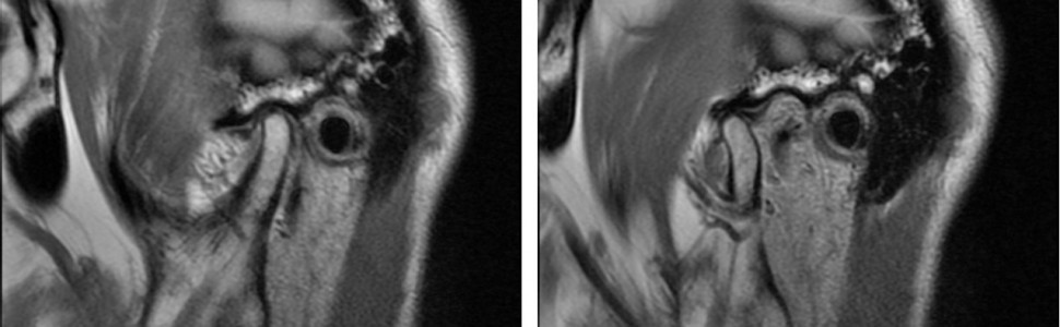
Imaging diagnostics of temporomandibular joints by cone-beam computed tomography and magnetic resonance imaging
Paweł Kozłowski, Beata Kozłowska, Edward Kijak
Streszczenie
Dysfunkcje stawów skroniowo-żuchwowych są często występującą grupą zaburzeń dotyczących układu stomatognatycznego. Ich objawy mogą być różnorodne, od bezbólowego zaburzenia toru prowadzenia żuchwy do trzasków i trzeszczeń podczas odwodzenia żuchwy, którym towarzyszą samoistne lub sprowokowane żuciem objawy bólowe w okolicy przedusznej. Współczesne metody obrazowania struktur wchodzących w skład stawów skroniowo-żuchwowych pozwalają na wczesne wykrycie zmian morfologicznych, a co za tym idzie – wczesne podjęcie leczenia i prognozowanie postępu zmian. Celem pracy jest porównanie dwóch metod obrazowania poszczególnych elementów stawów skroniowo-żuchwowych: stożkowej tomografii komputerowej (CBCT) i obrazowania metodą rezonansu magnetycznego (MRI).
Abstract
The temporomandibular joint (TMJ) dysfunctions represent a frequent group of disorders occurring in the stomatognathic system. Their symptoms may range from painless abnormalities of the mandibular path to snapping and crackling during mandibular abduction, which may be accompanied by either spontaneous or chewing pain in the pre-parotid region. Modern methods of imaging the structures that make up the temporomandibular joint allow early detection of morphological changes, enabling early treatment and prognosis of growth changes. The aim of this study is to compare two methods of imaging individual elements of the temporomandibular joint: cone-beam computed tomography (CBCT) and magnetic resonance imaging (MRI).
Hasła indeksowe: staw skroniowo-żuchwowy, stożkowa tomografia komputerowa, CBCT, obrazowanie metodą rezonansu magnetycznego, MRI
Key words: temporomandibular joint, cone-beam computed tomography, CBCT, magnetic resonance imaging, MRI
PIŚMIENNICTWO
- Tomasik E. i wsp.: Budowa anatomiczna stawu skroniowo-żuchwowego. Mag. Stomatol., 2009, 19, 3, 10-16.
- Farman A.G., Scarfe W.C.: The basics of maxillofacial cone beam computed tomography. Semin. Orthod., 2009, 15, 1, 2-13.
- De Vos W., Casselman J., Swennen G.R.: Cone-beam computerized tomography (CBCT) imaging of the oral and maxillofacial region. A systematic review of the literature. Int. J. Oral Maxillofac. Surg., 2009, 38, 6, 609-625.
- Librizzi Z.T. i wsp.: Cone-beam computed tomography to detect erosions of the temporomandibular joint. Effect of field of view and voxel size on diagnostic efficacy and effective dose. Am. J. Orthod. Dentofacial Orthop., 2011, 140, 1, e25-30.
- Palomo J.M., Rao P.S., Hans M.G.: Influence of CBCT exposure conditions on radiation dose. Oral Surg. Oral Med. Oral Pathol. Oral Radiol. Endod., 2008, 105, 6, 773-782.
- Kijak E., Lietz-Kijak D.: Asymetria twarzy w badaniach tomografii wolumetrycznej u pacjentów z zaburzeniami czynnościowymi układu ruchowego narządu żucia. Mag. Stomatol., 2018, 28, 5, 26-32.
- Ludlow J.B., Ivanovic M.: Comparative dosimetry of dental CBCT devices and 64-slice CT for oral and maxillofacial radiology. Oral Surg. Oral Med. Oral Pathol. Oral Radiol. Endod., 2008, 106, 1, 106-114.
- Faccioli N. i wsp.: Radiation dose saving through the use of cone-beam CT in hearing-impaired patients. Med., 2009, 114, 8, 1308-1318.
- Kijak E. i wsp.: Zmiany w strukturach kostnych stawów skroniowo-żuchwowych pacjentów z dysfunkcjami w diagnostyce CBCT. Mag. Stomatol., 2016, 26, 5, 32-38.
- Kijak E. i wsp.: Zastosowanie nowoczesnych technik radiologicznych w diagnostyce różnicowej dysfunkcji stawów skroniowo-żuchwowych na podstawie wybranych przypadków. Stomatol., 2014, 24, 5, 12-18.
- White S.C., Pharoah M.J.: Radiologia stomatologiczna. Wydawnictwo Czelej, Lublin 2002.
- Junhasavasdikul T. i wsp.: Developing a reference MRI database for temporomandibular joints in healthy children and adolescents. Radiol., 2018, 48, 8, 1113-1122.
- Pączkowski P.: Podstawy fizyczne i historia obrazowania metodą rezonansu magnetycznego. Wszechświat, 2012, 113, 292-302.
- Litko M. i wsp.: Correlation between the lateral pterygoid muscle attachment type and temporomandibular joint disc position in magnetic resonance imaging. Dentomaxillofac. Radiol., 2016, 45, 8, 2016-2029.
- Larheim T.A.: Role the magnetic resonance imaging in the clinical diagnosis of the temporomandibular joint. Cells Tissues Organs, 2005, 180, 1, 6-21.
- Litko M. i wsp.: Correlation between direction and severity of temporomandibular joint disc displacement and reduction ability during mouth opening. J. Oral Rehabil., 2017, 44, 12, 957-963.
- Roberts D. i wsp.: Temporomandibular joint. Magnetic resonance imaging. Radiology, 1985, 154, 829-830.
- Nitzan D.W.: The process of lubrication impairment and its involvement in temporomandibular joint disc displacement. A theoretical concept. J. Oral Maxillofac. Surg., 2001, 59, 1, 36-45.
- Manfredini D.: Etiopathogenesis of disc displacement of the temporomandibular joint. A review of the mechanisms. Indian J. Dent. Res., 2009, 20, 2, 212-221.
- Costen J.B.: Neuralgias and ear symptoms associated with disturbed function of the temporomandibular joint. JAMA, 1936, 107, 4, 252-255.
- Slaviček R.: The masticatory organ. GAMMA, Klosterneuburg 2006.
- Wilkinson T.M., Crowley C.M.: A histologic study retrodiscal tissues of the human temporomandibular joint in the open and closed position. J. Orofac. Pain., 1994, 8, 1, 7-17.
- Ogϋtcen-Toller M., Juniper R.P.: The development of the human lateral pterygoid muscle and the temporomandibular joint and related structures. A three-dimensional approach. Early Hum. Dev., 1994, 39, 1, 57-68.
- Nitzan D.W.: Intraarticular pressure in the functioning human temporomandibular joint and its alteration by uniform elevation of the occlusal plane. Oral Maxillofac. Surg., 1994, 52, 7, 671-679.
- Okeson J.P.: Leczenie dysfunkcji narządu żucia i zaburzeń zwarcia. Wydawnictwo Czelej, Lublin 2001.
- Alomar X. i wsp.: Anatomy of the temporomandibular joint. Sem. Ultrasound CT MRI, 2007, 28, 3, 170-183.
- Kondoh T. i wsp.: Prevalence of morphological changes in the surfaces of the temporomandibular joint disc associated with internal derangement. J. Oral Maxillofac. Surg., 1998, 56, 3, 339-343.
- Dergin G. i wsp.: Evaluating the correlation between the lateral pterygoid muscle attachment type and internal derangement of the temporomandibular joint with an emphasis on MR imaging findings. J. Craniomaxillofac. Surg., 2012, 40, 5, 459-463.
- Imanimoghaddam M., Madani A.S., Hashemi E.M.: The evaluation of lateral pterygoid muscle pathologic changes and insertion patterns in temporomandibular joints with or without disc displacement using magnetic resonance imaging. Int. J. Oral Maxillofac. Surg., 2013, 42, 9, 1116-1120.
- Antropoulou M. i wsp.: Variations of the attachment of the superior head of human lateral pterygoid muscle. J. Craniomaxillofac. Surg., 2013, 41, 6, e91-97.
- Pompei Filho H., Suazo G.I.C., Guimaraes A.S.: Prevalence of the third head lateral pterygoid muscle. A magnetic resonance image study. Int. J. Morphol., 2009, 27, 4, 1043-1046.
- Wilkinson T.M.: The relationship between the disk and lateral pterygoid muscle in the human temporomandibular joint. J. Prosthet. Dent., 1988, 60, 6, 715-724.
- Heylings D.J., Nielsen I.L., McNeill C.: Lateral pterygoid muscle and the temporomandibular disc. Orofac. Pain, 1995, 9, 1, 9-16.
- Mlosek K.: Radiologia stomatologiczna i szczękowo-twarzowa. Wydawnictwo Meddentpress, Warszawa 1995.
- Molina O.F. i wsp.: Oral jaw behaviors in TMD and bruxism. A comparison study by severity of bruxism. Cranio, 2001, 19, 2, 114-122.
- Manfredini D. i wsp.: Prevalence of bruxism in patients with different research diagnostic criteria for temporomandibular disorders (RDC/TMD) diagnoses. Cranio, 2003, 21, 4, 279-285.


