

Dostęp do tego artykułu jest płatny.
Zapraszamy do zakupu!
Po dokonaniu zakupu artykuł w postaci pliku PDF prześlemy bezpośrednio pod twój adres e-mail.
Torbiel zastoinowa lewej zatoki szczękowej o nietypowym przebiegu – opis leczenia chirurgicznego z porównaniem metod leczenia
Left maxillary sinus retention cyst with atypical symptoms – surgical case report with treatment methods comparison
Mateusz Halama, Kamil Nelke, Maciej Stagraczyński, Radosław Jadach
Streszczenie
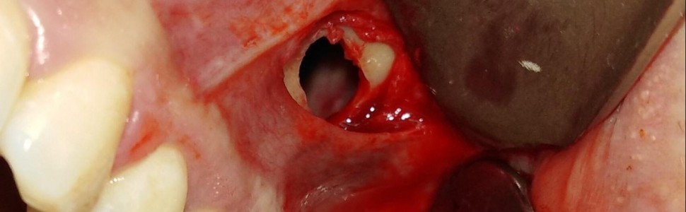
Celem niniejszej pracy jest prezentacja przypadku leczenia operacyjnego zmiany zatoki szczękowej lewej o charakterze torbieli zastoinowej. Większość zmian tego typu nie wymaga leczenia operacyjnego, o ile nie daje objawów patologicznych. Niektóre klasyfikacje wyróżniają dwa podtypy zmiany: śluzową i surowiczą. Przyjmuje się, że zmiany nieprzekraczające 50% objętości zatoki szczękowej bardzo rzadko dają objawy kliniczne i zazwyczaj nie wymagają leczenia chirurgicznego. W szczególnych przypadkach nietypowych dolegliwości u pacjentów należy rozszerzyć diagnostykę oraz zweryfikować słuszność dalszego leczenia chirurgicznego, endodontycznego, a może i skojarzonego laryngologiczno-stomatologicznego. W pracy przedstawiono opis przypadku pacjentki, która po próbie leczenia farmakologicznego i stomatologicznego (leczenie endodontyczne zęba 26) wymagała leczenia chirurgicznego metodą zmodyfikowanej operacji Caldwella-Luca.
Abstract
The aim of this study is to present a case of surgical treatment of a lesion in the left maxillary sinus in the form of a retention cyst of the maxillary sinus. Most lesions of this type do not require surgical treatment, unless they show pathological symptoms. Some authors discuss two types of lesions: mucous and serous. It is assumed that lesions not exceeding 50% of the maxillary sinus volume very rarely give rise to clinical symptoms and usually do not require surgery. In some cases when atypical symptoms occur, in some patients it is necessary to improve further diagnostic steps and evaluate the accuracy of other surgical, dental or perhaps combined laryngological-dental approaches. The paper presents a description of a case of a patient who, after trying pharmacological and dental treatment (endodontic treatment of tooth 26), required surgical treatment using a modified Caldwell-Luc operation.
Hasła indeksowe: torbiel zastoinowa, zatoka szczękowa, operacja Caldwella-Luca, bóle zatoki, objaw zatkanego nosa
Key words: mucous retention cyst, maxillary sinus, Caldwell-Luc procedure, sinus pains, swollen nose syndrome
Piśmiennictwo
- Bal M, Saltürk Z, Bal GC i wsp. Mucous retention cysts in the paranasal sinuses. A retrospective study. Otolaryngol J. 2016; 6: 1-6.
- Gordts F, Clement PA, Buisseret T. Prevalence of paranasal sinus abnormalities on MRI in non-ENT population. Acta Otorhinolaryngol Belg. 1996; 50(3): 167-170.
- Santosh U. Maxillary sinus retention cyst. An unusual presentation. Gujarat J Otorhinol Head Neck Surg. 2010; 7(1): 36-37.
- Gothberg KA, Little JW, King DR i wsp. A clinical study of cysts arising from mucosa of the maxillary sinus. Oral Surg Oral Med Oral Pathol. 1976; 41(1): 52-58.
- Kaczmarzyk T. Torbiele obszaru szczękowo-twarzowego. Warszawa: Wydawnictwo Kwintesencja; 2015.
- Gaillard F, Ashraf A. Paranasal sinus mucocele. Online: https://radiopaedia.org/articles/paranasal-sinus-mucocele-1 [dostęp: 11.02.2020].
- Meer S, Altini M. Cysts and pseudocysts of the maxillary antrum revisited. SADJ. 2006; 61(1): 10-13.
- Lacin N, Tatar B. Evaluation of the frequency of mucous retention cysts in the maxillary sinus in a Turkish population using cone-beam computed tomography. Makara J Health Res. 2019; 23(2): 68-71.
- Hoang JK, Smith EC, Barboriak DP. Ruptured maxillary retention cyst. Cause of unilateral rhinorrhea after trauma. AJNR Am J Neuroradiol. 2009; 30(6): 1121-1122.
- Łasiński W. Anatomia głowy dla stomatologów. Warszawa: Państwowy Zakład Wydawnictw Lekarskich; 1993.
- Yeung AWK, Tanaka R, Khong PL i wsp. Frequency, location, and association with dental pathology of mucous retention cysts in the maxillary sinus. A radiographic study using cone beam computed tomography (CBCT). Clin Oral Investig. 2018; 22(3): 1175-1183.
- Bhattacharyya N. Do maxillary sinus retention cysts reflect obstructive sinus phenomena? Arch Otolaryngol Head Neck Surg. 2000; 126(11): 1369-1371.
- Gonçales ES, Gonçales AGB, da Silva Lima E i wsp. Symptomatic mucous retention cysts of the maxillary sinus. Case report. RSBO. 2015; 12(2): 233-237.
- Albu S. Symptomatic maxillary sinus retention cysts. Should they be removed? Laryngoscope. 2010; 120(9): 1904-1909.
- Hadar T, Shvero J, Nageris BI i wsp. Mucus retention cyst of the maxillary sinus. The endoscopic approach. Br J Oral Maxillofac Surg. 2000; 38(3): 227-229.
- Kasamatsu A, Fukumoto C, Higo M i wsp. Treatment of an extensive maxillary cyst using nasal airway and balloon catheter devices. Case Rep Dent. 2014; 2014: 216828.
- Nilesh K, Dadhich A. Unusually large radicular cyst presenting in the maxillary sinus. BMJ Case Rep. 2020; 13(9): e236582.
- Mattos RG, Egas LS, Oliveira A i wsp. Mucous retention cyst in maxillary sinus with expansion of maxillary tuberosity. Case Report. J Oral Diag. 2018; 03: e20180003.


