

Dostęp do tego artykułu jest płatny.
Zapraszamy do zakupu!
Cena: 12.50 PLN (z VAT)
Kup artykuł
Po dokonaniu zakupu artykuł w postaci pliku PDF prześlemy bezpośrednio pod twój adres e-mail.
MS 2020; 6: 50-54.
Jacek Matys
Streszczenie
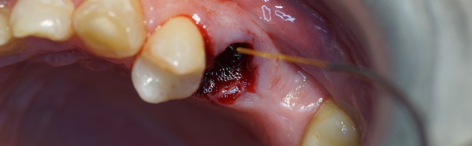
W pracy omówiono zasady wytwarzania skrzepu w zębodołach poekstrakcyjnych za pomocą lasera diodowego o długości fali 980 nm u pacjentów zażywających przewlekle leki przeciwzakrzepowe. Laser o długości fali 980 nm jest skutecznym narzędziem pozwalającym na stabilizację skrzepu krwi w zębodole.
Abstract
The paper discusses the principles of blood clot formation in extraction socket using a diode laser with a wavelength of 980 nm in patients taking anticoagulants. The 980 nm laser is an effective tool that helps stabilize the blood clot in the extraction socket.
Hasła indeksowe: laser diodowy, koagulacja, hemostaza naczyniowa
Key words: diode laser, coagulation, vascular hemostasis
Piśmiennictwo
1. Ruan Y, Guo Y, Zheng Y i wsp. Cardiovascular disease (CVD) and associated risk factors among older adults in six low-and middle-income countries. Results from SAGE Wave 1. BMC Public Health. 2018; 18(1): 778.
2. Cayla G, Morange PE, Chambost H i wsp. Management of cardiovascular disease in haemophilia. Thromb Res. 2013; 132(1): 8-14.
3. Najafzadeh M, Schneeweiss S, Choudhry NK i wsp. General population vs. patient preferences in anticoagulant therapy. A discrete choice experiment. Patient. 2019; 12(2): 235-246.
4. Sidelmann JJ, Gram J, Jespersen J i wsp. Fibrin clot formation and lysis. Basic mechanisms. Semin Thromb Hemost. 2000; 26(6): 605-618.
5. Matys J. Leczenie zmian naczyniowych za pomocą lasera diodowego o długości fali 980 nm. Med Trib Stomatol. 2019; 3.
6. Matys J, Jaszczak E, Flieger R i wsp. Effect of ozone and diode laser (635 nm) in reducing orthodontic pain in the maxillary arch. A randomized clinical controlled trial. Lasers Med Sci. 2020; 35(2): 487-496.
7. Matys J, Świder K, Flieger R. Laser instant implant impression method. A case presentation. Dent Med Probl. 2017; 54(1).
8. Matys J, Dominiak M. Assessment of pain when uncovering implants with Er:YAG laser or scalpel for second stage surgery. Adv Clin Exp Med. 2016; 25(6): 1179-1184.
9. Milonni PW, Eberly JH. Laser physics. Wyd. 2. Hoboken: Wiley; 2010, s. 38-40.
10. de Freitas PM, Simoes A. Lasers in dentistry. Guide for clinical practice. Saint Louis: Mosby; 2015.
11. Matys J, Dominiak M. Ocena bólu podczas odsłaniania implantów za pomocą lasera erbowo-yagowego. Implantol Stomatol. 2014; 5(2): 52-54.
12. Ishikawa I, Aoki A, Takasaki AA. Potential applications of Erbium:YAG laser in periodontics. J Periodontal Res. 2004; 39(4): 275-285.
13. Sułek J. Możliwości wykorzystania laserów w protetyce stomatologicznej. Porad Stomatol. 2010; 10(4): 139-143.
14. Wendt-Nordahl G, Huckele S, Honeck i wsp. 980-nm Diode laser: a novel laser technology for vaporization of the prostate. European urology. 2007; 52(6):1723-8.
Streszczenie
W pracy omówiono zasady wytwarzania skrzepu w zębodołach poekstrakcyjnych za pomocą lasera diodowego o długości fali 980 nm u pacjentów zażywających przewlekle leki przeciwzakrzepowe. Laser o długości fali 980 nm jest skutecznym narzędziem pozwalającym na stabilizację skrzepu krwi w zębodole.
Abstract
The paper discusses the principles of blood clot formation in extraction socket using a diode laser with a wavelength of 980 nm in patients taking anticoagulants. The 980 nm laser is an effective tool that helps stabilize the blood clot in the extraction socket.
Hasła indeksowe: laser diodowy, koagulacja, hemostaza naczyniowa
Key words: diode laser, coagulation, vascular hemostasis
Piśmiennictwo
1. Ruan Y, Guo Y, Zheng Y i wsp. Cardiovascular disease (CVD) and associated risk factors among older adults in six low-and middle-income countries. Results from SAGE Wave 1. BMC Public Health. 2018; 18(1): 778.
2. Cayla G, Morange PE, Chambost H i wsp. Management of cardiovascular disease in haemophilia. Thromb Res. 2013; 132(1): 8-14.
3. Najafzadeh M, Schneeweiss S, Choudhry NK i wsp. General population vs. patient preferences in anticoagulant therapy. A discrete choice experiment. Patient. 2019; 12(2): 235-246.
4. Sidelmann JJ, Gram J, Jespersen J i wsp. Fibrin clot formation and lysis. Basic mechanisms. Semin Thromb Hemost. 2000; 26(6): 605-618.
5. Matys J. Leczenie zmian naczyniowych za pomocą lasera diodowego o długości fali 980 nm. Med Trib Stomatol. 2019; 3.
6. Matys J, Jaszczak E, Flieger R i wsp. Effect of ozone and diode laser (635 nm) in reducing orthodontic pain in the maxillary arch. A randomized clinical controlled trial. Lasers Med Sci. 2020; 35(2): 487-496.
7. Matys J, Świder K, Flieger R. Laser instant implant impression method. A case presentation. Dent Med Probl. 2017; 54(1).
8. Matys J, Dominiak M. Assessment of pain when uncovering implants with Er:YAG laser or scalpel for second stage surgery. Adv Clin Exp Med. 2016; 25(6): 1179-1184.
9. Milonni PW, Eberly JH. Laser physics. Wyd. 2. Hoboken: Wiley; 2010, s. 38-40.
10. de Freitas PM, Simoes A. Lasers in dentistry. Guide for clinical practice. Saint Louis: Mosby; 2015.
11. Matys J, Dominiak M. Ocena bólu podczas odsłaniania implantów za pomocą lasera erbowo-yagowego. Implantol Stomatol. 2014; 5(2): 52-54.
12. Ishikawa I, Aoki A, Takasaki AA. Potential applications of Erbium:YAG laser in periodontics. J Periodontal Res. 2004; 39(4): 275-285.
13. Sułek J. Możliwości wykorzystania laserów w protetyce stomatologicznej. Porad Stomatol. 2010; 10(4): 139-143.
14. Wendt-Nordahl G, Huckele S, Honeck i wsp. 980-nm Diode laser: a novel laser technology for vaporization of the prostate. European urology. 2007; 52(6):1723-8.


