

Dostęp do tego artykułu jest płatny.
Zapraszamy do zakupu!
Cena: 12.50 PLN (z VAT)
Kup artykuł
Po dokonaniu zakupu artykuł w postaci pliku PDF prześlemy bezpośrednio pod twój adres e-mail.
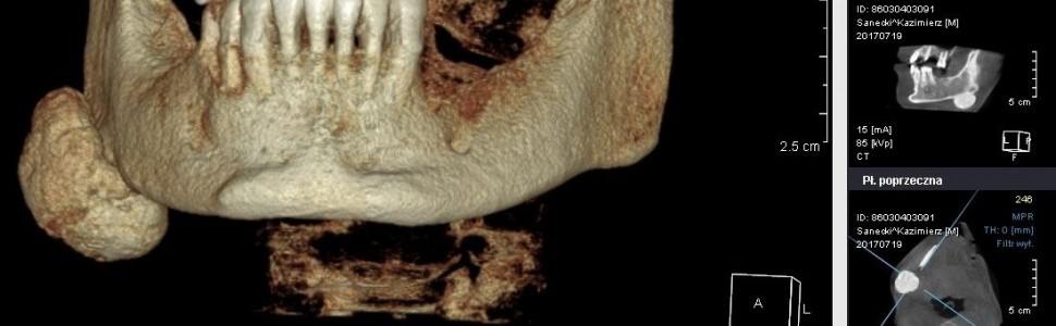
Mandibular osteoma – case study
Mansur Rahnama, Dagmara Jenda, Joanna Jakiel, Marcin Kornet, Michał ŁobaczStreszczenie
Kostniaki to łagodne guzy złożone z dojrzałej kości zbitej i gąbczastej. Dzielą się na obwodowe, centralne i pozaszkieletowe. Kostniaki obwodowe biorą swój początek z okostnej i wzrastają na powierzchni kości, centralne rozwijają się wewnątrz kości ze szpiku kostnego, a pozaszkieletowe zwykle obserwuje się wewnątrz mięśni. Kostniaki wykrywa się najczęściej w kościach twarzy i czaszki. Obecność kostniaków mnogich może być związana z zespołem Gardnera. Zmiany te są przeważnie bezbolesne, a wznowy po ich usunięciu występują niezwykle rzadko. Autorzy pracy prezentują przypadek 31-letniego pacjenta leczonego w Zakładzie Chirurgii Stomatologicznej z powodu kostniaka obwodowego, zlokalizowanego w pobliżu kąta żuchwy strony prawej.
Abstract
Osteoma is a benign tumour composed of compact bone and cancellous bone. They are divided into peripheral, central and extra-skeletal. Peripheral osteoma originates from the periosteum and grows on the surface of the bone. Central osteoma develops in an intraosseous manner out of bone marrow. Whereas, extra-skeletal osteomas are often defined as intramuscular. Osteoma occurs most frequently in facial and skull bones. The incidence of multiple osteomas may be connected with Gardner’s syndrome. The lesions are usually painless and post-removal recurrences are extremely rare. The authors of the paper present the case of a 31-year-old patient, treated in the Department of Oral Surgery, suffering from peripheral osteoma located near the right mandibular angle.
Hasła indeksowe: kostniak, CBCT
Key words: osteoma, CBCT
PIŚMIENNICTWO
1. Horikawa F.K. i wsp.: Peripheral osteoma of the maxillofacial region. A study of 10 cases. Braz. J. Otorhinolaryngol., 2012, 78, 5, 5, 38-43.
2. Gumusok M. i wsp.: Peripheral osteoma of the mandible. A case report. J. Istanb. Univ. Fac. Dent., 2015, 49, 1, 47-50.
3. Viswanatha B.: Maxillary sinus osteoma. Two cases and review of the literature. Acta Otorhinolaryngol. Ital., 2012, 32, 3, 202-205.
4. Borumandi F. i wsp.: Maxillary sinus osteoma. From incidental finding to surgical management. J. Oral. Maxillofac. Pathol., 2013, 17, 2, 318.
5. Nah K.S.: Osteomas of the craniofacial region. Imaging Sci. Dent., 2011, 41, 3, 107-113.
6. Janas A., Osica P.: Mnogie kostniaki zewnątrzkostne. Dental Forum, 2015, 43, 1, 41-44.
7. Tarsitano A., Marchetti C.: Unusual presentation of obstructive sleep apnoea syndrome due to a giant mandible osteoma. Case report and literature review. Acta Otorhinolaryngol. Ital., 2013, 33, 1, 63-66.
8. De Souza N.T. i wsp.: An unusual osteoma in the mandibular condyle and the successful replacement of the temporomandibular joint with a custom-made prosthesis. A case report. BMC Res. Notes, 2017, 10, 1, 727.
9. Woldenberg Y., Nash M., Bodner L.: Peripheral osteoma of the maxillofacial region. Diagnosis and management. A study of 14 cases. Med. Oral. Patol. Oral. Cir. Bucal, 2005, 10 Suppl. 2, E139-142.
10. D’Amato S. i wsp.: Piezoelectric bone surgery in the treatment of an osteoma associated with an impacted inferior third molar. A case report. Clin. Cases Miner. Bone Metab., 2014, 11, 1, 73-76.


